
 | ||
Gastrointestinal is an adjective meaning of or pertaining to the stomach and intestines. A tract is a collection of related anatomic structures or a series of connected body organs.
Contents
- Structure
- Upper gastrointestinal tract
- Lower gastrointestinal tract
- Development
- Histology
- Function
- Immune function
- Intestinal microbiota
- Detoxification and drug metabolism
- Diseases
- Symptoms
- Treatment
- Imaging
- Other related diseases
- Uses of animal guts
- Other animals
- References
The gastrointestinal tract (digestive tract, GI tract, GIT, gut, or alimentary canal) is an organ system within humans and other animals which takes in food, digests it to extract and absorb energy and nutrients, and expels the remaining waste as feces and urine. The mouth, oesophagus, stomach, and intestines are part of the human alimentary canal.
All bilaterians have a gastrointestinal tract, also called a gut or an alimentary canal. This is a tube that transfers food to the organs of digestion. In large bilaterians, the gastrointestinal tract generally also has an exit, the anus, by which the animal disposes of feces (solid wastes). Some small bilaterians have no anus and dispose of solid wastes by other means (for example, through the mouth).
The gastrointestinal tract contains thousands of different bacteria in their gut flora.
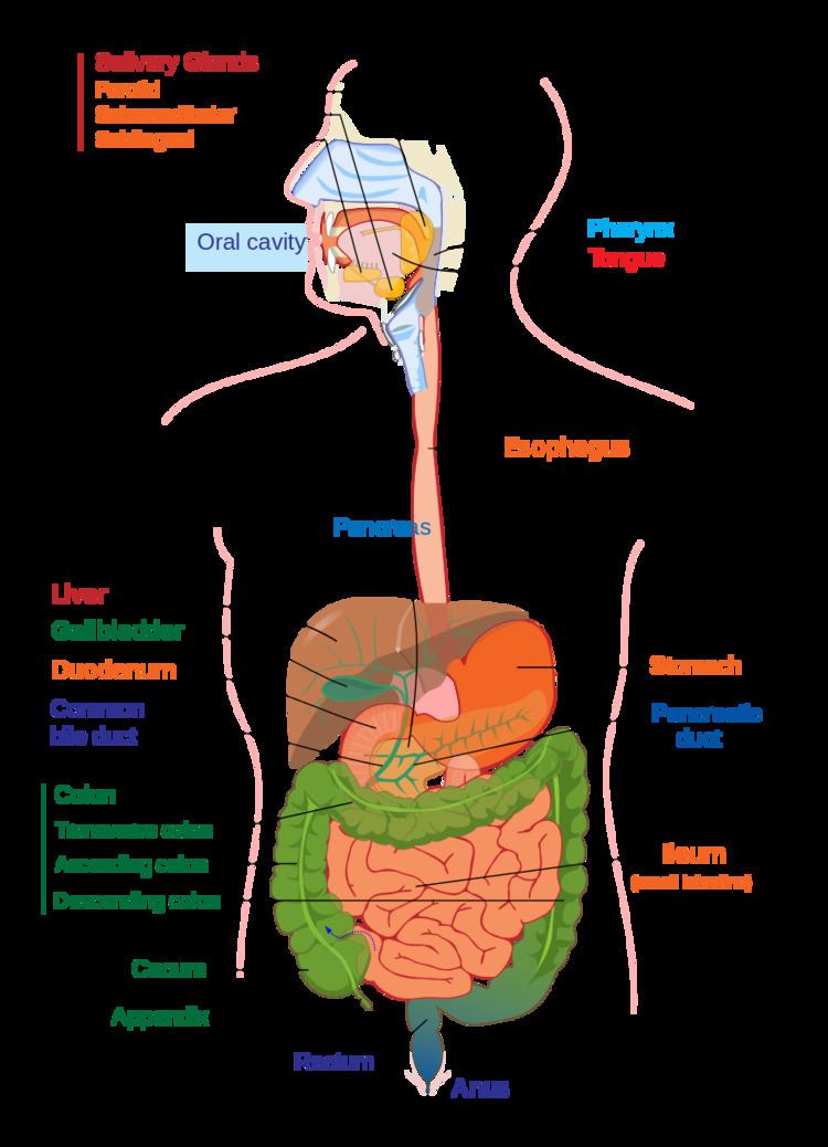
The human gastrointestinal tract consists of the esophagus, stomach, and intestines, and is divided into the upper and lower gastrointestinal tracts. The GI tract includes all structures between the mouth and the anus, forming a continuous passageway that includes the main organs of digestion, namely, the stomach, small intestine, and large intestine. In contrast, the human digestive system comprises the gastrointestinal tract plus the accessory organs of digestion (the tongue, salivary glands, pancreas, liver, and gallbladder). The tract may also be divided into foregut, midgut, and hindgut, reflecting the embryological origin of each segment.
The whole human GI tract is about nine metres (30 feet) long at autopsy. It is considerably shorter in the living body because the intestines, which are tubes of smooth muscle tissue, maintain constant muscle tone, somewhat like a slinky that maintains itself in a halfway-tense state but can relax in spots to allow for local distention, peristalsis, and so on.
The GI tract releases hormones from enzymes to help regulate the digestive process. These hormones, including gastrin, secretin, cholecystokinin, and ghrelin, are mediated through either intracrine or autocrine mechanisms, indicating that the cells releasing these hormones are conserved structures throughout evolution.
Structure
The structure and function can be described both as gross anatomy and as microscopic anatomy or histology. The tract itself is divided into upper and lower tracts, and the intestines small and large parts.
Upper gastrointestinal tract
The upper gastrointestinal tract consists of the buccal cavity, pharynx, esophagus, stomach, and duodenum. The exact demarcation between the upper and lower tracts is the suspensory muscle of the duodenum. This delineates the embryonic borders between the foregut and midgut, and is also the division commonly used by clinicians to describe gastrointestinal bleeding as being of either "upper" or "lower" origin. Upon dissection, the duodenum may appear to be a unified organ, but it is divided into four segments based upon function, location, and internal anatomy. The four segments of the duodenum are as follows (starting at the stomach, and moving toward the jejunum): bulb, descending, horizontal, and ascending. The suspensory muscle attaches the superior border of the ascending duodenum to the diaphragm.
The suspensory muscle is an important anatomical landmark which shows the formal division between the duodenum and the jejunum, the first and second parts of the small intestine, respectively. This is a thin muscle which is derived from the embryonic mesoderm.
Lower gastrointestinal tract
The lower gastrointestinal tract includes most of the small intestine and all of the large intestine. In human anatomy, the intestine (bowel, or gut) is the segment of the gastrointestinal tract extending from the pyloric sphincter of the stomach to the anus and, in humans and other mammals, consists of two segments, the small intestine and the large intestine. In humans, the small intestine is further subdivided into the duodenum, jejunum and ileum while the large intestine is subdivided into the cecum, colon, rectum, and anal canal.
Small intestine
The small intestine begins at the duodenum, which receives food from the stomach. It is a tubular structure, usually between 6 and 7 m long. The area of the human, adult small intestinal mucosa is about 30 m2. Its main function is to absorb the products of digestion (including carbohydrates, proteins, lipids, and vitamins) into the bloodstream. It has three major divisions:
- Duodenum: A short structure (about 20–25 cm long) which receives chyme from the stomach, together with pancreatic juice containing digestive enzymes and bile from the gall bladder. The digestive enzymes break down proteins, and bile emulsifies fats into micelles. The duodenum contains Brunner's glands, which produce a mucus-rich alkaline secretion containing bicarbonate. These secretions, in combination with bicarbonate from the pancreas, neutralizes the stomach acids contained in the chyme.
- Jejunum: This is the midsection of the small intestine, connecting the duodenum to the ileum. It is about 2.5 m long, and contains the circular folds, and villi that increase its surface area. Products of digestion (sugars, amino acids, and fatty acids) are absorbed into the bloodstream here.
- Ileum: The final section of the small intestine. It is about 3 m long, and contains villi similar to the jejunum. It absorbs mainly vitamin B12 and bile acids, as well as any other remaining nutrients.
Large intestine
The large intestine also called the colon, consists of the cecum, rectum, and anal canal. It also includes the appendix, which is attached to the cecum. The colon is further divided into:
- Cecum (first portion of the colon) and appendix
- Ascending colon (ascending in the back wall of the abdomen)
- Right colic flexure (flexed portion of the ascending and transverse colon apparent to the liver)
- Transverse colon (passing below the diaphragm)
- Left colic flexure (flexed portion of the transverse and descending colon apparent to the spleen)
- Descending colon (descending down the left side of the abdomen)
- Sigmoid colon (a loop of the colon closest to the rectum)
- Rectum
- Anus
The main function of the large intestine is to absorb water. The area of the large intestinal mucosa of an adult human is about 2 m2.
Development
The gut is an endoderm-derived structure. At approximately the sixteenth day of human development, the embryo begins to fold ventrally (with the embryo's ventral surface becoming concave) in two directions: the sides of the embryo fold in on each other and the head and tail fold toward one another. The result is that a piece of the yolk sac, an endoderm-lined structure in contact with the ventral aspect of the embryo, begins to be pinched off to become the primitive gut. The yolk sac remains connected to the gut tube via the vitelline duct. Usually this structure regresses during development; in cases where it does not, it is known as Meckel's diverticulum.
During fetal life, the primitive gut is gradually patterned into three segments: foregut, midgut, and hindgut. Although these terms are often used in reference to segments of the primitive gut, they are also used regularly to describe regions of the definitive gut as well.
Each segment of the gut is further specified and gives rise to specific gut and gut-related structures in later development. Components derived from the gut proper, including the stomach and colon, develop as swellings or dilatations in the cells of the primitive gut. In contrast, gut-related derivatives — that is, those structures that derive from the primitive gut but are not part of the gut proper, in general develop as out-pouchings of the primitive gut. The blood vessels supplying these structures remain constant throughout development.
Histology
The gastrointestinal tract has a form of general histology with some differences that reflect the specialization in functional anatomy. The GI tract can be divided into four concentric layers in the following order:
Mucosa
The mucosa is the innermost layer of the gastrointestinal tract. that is surrounding the lumen, or open space within the tube. This layer comes in direct contact with digested food (chyme). The mucosa is made up of:
The mucosae are highly specialized in each organ of the gastrointestinal tract to deal with the different conditions. The most variation is seen in the epithelium.
Submucosa
The submucosa consists of a dense irregular layer of connective tissue with large blood vessels, lymphatics, and nerves branching into the mucosa and muscularis externa. It contains the submucosal plexus, an enteric nervous plexus, situated on the inner surface of the muscularis externa.
Muscular layer
The muscular layer consists of an inner circular layer and a longitudinal outer layer. The circular layer prevents food from traveling backward and the longitudinal layer shortens the tract. The layers are not truly longitudinal or circular, rather the layers of muscle are helical with different pitches. The inner circular is helical with a steep pitch and the outer longitudinal is helical with a much shallower pitch.
Between the two muscle layers is the myenteric plexus. This controls peristalsis. Activity is initiated by the pacemaker cells, (myenteric interstitial cells of Cajal). The gut has intrinsic peristaltic activity (basal electrical rhythm) due to its self-contained enteric nervous system. The rate can be modulated by the rest of the autonomic nervous system.
The coordinated contractions of these layers is called peristalsis and propels the food through the tract. Food in the GI tract is called a bolus (ball of food) from the mouth down to the stomach. After the stomach, the food is partially digested and semi-liquid, and is referred to as chyme. In the large intestine the remaining semi-solid substance is referred to as faeces.
Adventitia and serosa
The outermost layer of the gastrointestinal tract consists of several layers of connective tissue.
Intraperitoneal parts of the GI tract are covered with serosa. These include most of the stomach, first part of the duodenum, all of the small intestine, caecum and appendix, transverse colon, sigmoid colon and rectum. In these sections of the gut there is clear boundary between the gut and the surrounding tissue. These parts of the tract have a mesentery.
Retroperitoneal parts are covered with adventitia. They blend into the surrounding tissue and are fixed in position. For example, the retroperitoneal section of the duodenum usually passes through the transpyloric plane. These include the esophagus, pylorus of the stomach, distal duodenum, ascending colon, descending colon and anal canal. In addition, the oral cavity has adventitia.
Function
The time taken for food or other ingested objects to transit through the gastrointestinal tract varies depending on many factors, but roughly, it takes less than an hour after a meal for 50% of stomach contents to empty into the intestines while total emptying takes around 2 hours. Subsequently, 50% emptying of the small intestine takes between 1 and 2 hours. Finally, transit through the colon takes 12 to 50 hours with wide variation between individuals.
Immune function
Immune barrier
The gastrointestinal tract forms an important part of the immune system. The surface area of the digestive tract is estimated to be about 32 square meters, or about half a badminton court. With such a large exposure (more than three times larger than the exposed surface of the skin), these immune components function to prevent pathogens from entering the blood and lymph circulatory systems. Fundamental components of this protection are provided by the intestinal mucosal barrier which is composed of physical, biochemical, and immune elements elaborated by the intestinal mucosa. Microorganisms also are kept at bay by an extensive immune system comprising the gut-associated lymphoid tissue (GALT)
There are additional factors contributing to protection from pathogen invasion. For example, low pH (ranging from 1 to 4) of the stomach is fatal for many microorganisms that enter it. Similarly, mucus (containing IgA antibodies) neutralizes many pathogenic microorganisms. Other factors in the GI tract contribution to immune function include enzymes secreted in the saliva and bile.
Immune system homeostasis
Beneficial bacteria also can contribute to the gastrointestinal system homeostasis. A case in point is the relationship between human gut and Clostridia, one of the most predominant bacterial groups in the gastrointestinal tract. Clostridia play an important role influencing the dynamics of our immune system in the gut. It has been demonstrated that the intake of a high fiber diet could be the responsible for the induction of Treg cells. This is due to the production of short-chain fatty acids during the fermentation of plant derived nutrients such as butyrate and propionate. Basically, the butyrate induces the differentiation of Treg cells by enhancing histone H3 acetylation in the promoter and conserved non-coding sequence regions of the Foxp3 locus, and thus regulating the T cells, having as a result the reduction of the inflammatory response and allergies.
Intestinal microbiota
The large intestine hosts several kinds of bacteria that can deal with molecules that the human body cannot otherwise break down. This is an example of symbiosis. These bacteria also account for the production of gases at host-pathogen interface, inside our intestine(this gas is released as flatulence when eliminated through the anus). However the large intestine is mainly concerned with the absorption of water from digested material (which is regulated by the hypothalamus) and the re absorption of sodium, as well as any nutrients that may have escaped primary digestion in the ileum.
Health-enhancing intestinal bacteria of the gut flora serve to prevent the overgrowth of potentially harmful bacteria in the gut. These two types of bacteria compete for space and "food," as there are limited resources within the intestinal tract. A ratio of 80-85% beneficial to 15–20% potentially harmful bacteria generally is considered normal within the intestines.
Detoxification and drug metabolism
Enzymes such as CYP3A4, along with the antiporter activities, are also instrumental in the intestine's role of drug metabolism in the detoxification of antigens and xenobiotics.
Diseases
There are many diseases and conditions that can affect the gastrointestinal system, including infections, inflammation and cancer.
Various pathogens can cause gastroenteritis an inflammation of the stomach and small intestine. These can include those organisms that cause foodborne illnesses. Gastroenteritis is the most common disease of the GI tract.
Diverticular disease is a condition that is very common in older people in industrialized countries. It usually affects the large intestine but has been known to affect the small intestine as well. Diverticulosis occurs when pouches form on the intestinal wall. Once the pouches become inflamed it is known as diverticulitis.
Inflammatory bowel disease is an inflammatory condition affecting the bowel walls, and includes the subtypes Crohn's disease and ulcerative colitis. While Crohn's can affect the entire gastrointestinal tract, ulcerative colitis is limited to the large intestine. Crohn's disease is widely regarded as an autoimmune disease. Although ulcerative colitis is often treated as though it were an autoimmune disease, there is no consensus that it actually is such.
Functional gastrointestinal disorders the most common of which is irritable bowel syndrome. Functional constipation and chronic functional abdominal pain are other functional disorders of the intestine that have physiological causes, but do not have identifiable structural, chemical, or infectious pathologies.
Symptoms
Several symptoms are used to indicate problems with the gastrointestinal tract:
Treatment
Gastrointestinal surgery can often be performed in the outpatient setting. In the United States in 2012, operations on the digestive system accounted for 3 of the 25 most common ambulatory surgery procedures and constituted 9.1 percent of all outpatient ambulatory surgeries.
Imaging
Various methods of imaging the gastrointestinal tract include the upper and lower gastrointestinal series:
Other related diseases
Uses of animal guts
Animal intestines have multiple uses. From each species of livestock that is a source of milk, a corresponding rennet is obtained from the intestines of milk-fed calves. Pig and calf intestines are eaten, and pig intestines are used as sausage casings. Calf intestines supply calf-intestinal alkaline phosphatase (CIP), and are used to make goldbeater's skin. Other uses are:
Other animals
Many birds and other animals have a specialised stomach in the digestive tract called a gizzard used for grinding up food.
Another feature not found in the human but found in a range of other animals is the crop. In birds this is found as a pouch alongside the esophagus.
Other animals including amphibians, birds, reptiles, and egg-laying mammals have a major difference in their GI tract in that it ends in a cloaca and not an anus.