
Latin Canalis analis | ||
 | ||
Nerve autonomic inferior hypogastric plexus (above pectinate line) and somatic inferior rectal nerves (below line) Lymph Superficial inguinal lymph node (below pectinate line) and internal iliac lymph nodes (above line) | ||
The anal canal is the terminal part of the large intestine. It is situated between the rectum and anus, below the level of the pelvic diaphragm. It lies in the anal triangle of perineum in between the right and left ischioanal fossa.
Contents
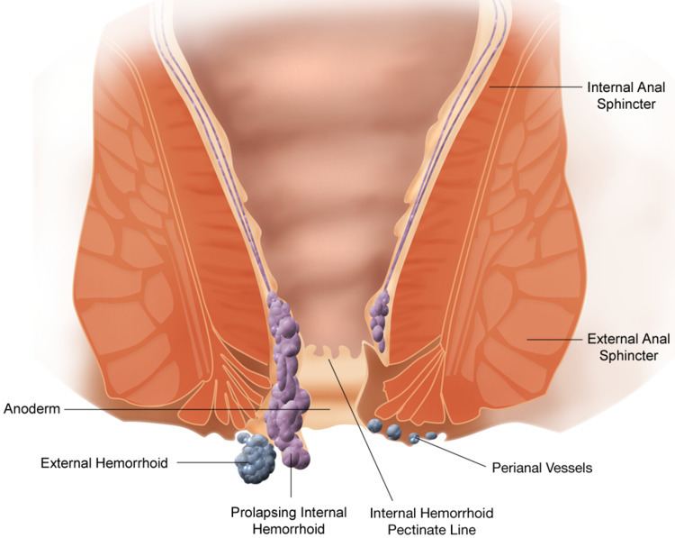
The anal canal is divided into three parts. The zona columnaris is the upper half of the canal and is lined by simple columnar epithelium. The lower half of the anal canal, below the pectinate line, is divided into two zones separated by Hilton's white line. The two parts are the zona hemorrhagica and zona cutanea, lined by stratified squamous non-keratinized and stratified squamous keratinized, respectively.
In humans it is approximately 2.5 to 4 cm long, extending from the anorectal junction to the anus. It is directed downwards and backwards. It is surrounded by inner involuntary and outer voluntary sphincters which keep the lumen closed in the form of an anteroposterior slit.
It is differentiated from the rectum by the transition of the internal surface from endodermal to skinlike ectodermal tissue.
Structure
The anal canal is divided into two unequal sections, upper and lower.
A whitish line called Hilton's white line or pecten of Robert Austin Stroud indicates the junction between keratinized stratified squamous epithelium and unkeratinized stratified squamous epithelium.
The anal verge is the distal end of the anal canal, forming a transitional zone between the epithelium of the anal canal and the perianal skin. It should not be confused with the "pectinate line".