
ICD-9-CM 562 MedlinePlus 000257 | ICD-10 K57 DiseasesDB 3876 eMedicine med/578 | |
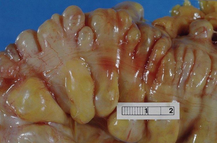 | ||
Diverticulitis is a digestive disease in which pouches within the large bowel wall become inflamed. Symptoms typically include lower abdominal pain of a sudden onset. Onset however may also occur over a few days. In North America and Europe pain is usually on the left side, while in Asia it is often on the right. There may also be fever, nausea, diarrhea or constipation, or blood in the stool. Repeated attacks may occur.
Contents
- Signs and symptoms
- Causes
- Diet
- Pathophysiology
- Diagnosis
- Differential diagnosis
- Treatment
- Antibiotics
- Surgery
- Complications
- Epidemiology
- References
The cause is uncertain. Risk factors may include obesity, lack of exercise, smoking, a family history of the disease, and nonsteroidal anti-inflammatory drugs (NSAIDs). The role of dietary fibre is unclear. Having pouches in the large intestine that are not inflamed is known as diverticulosis. Inflammation occurs in between 10% and 25% at some point in time and is due to a bacterial infection. Diagnosis may be made by blood tests, CT scan, colonoscopy, or lower gastrointestinal series. The differential diagnosis includes irritable bowel syndrome.
While avoiding nuts and seeds has historically been recommended, an association between eating these foods and diverticulitis has not been found, and therefore, avoiding these foods is no longer recommended. Mesalazine and rifaximin appear useful for preventing attacks in those with diverticulosis. For mild diverticulitis, antibiotics by mouth and a liquid diet is recommended. For severe cases intravenous antibiotics, hospital admission, and complete bowel rest may be recommended. Probiotics are of unclear use. Complications such as abscess formation, fistula formation, and perforation of the colon may require surgery.
The disease is common in the Western world and uncommon in Africa and Asia. In the Western world about 35% of people have diverticulosis while it affects less than 1% of those in rural Africa. The disease becomes more frequent with age being particularly common in those over the age of 50. It has also become more common in all parts of the world. In 2003 in Europe it resulted in about 13,000 deaths. Costs associated with diverticular disease are around $2.4 billion a year in the United States as of 2013.
Signs and symptoms
People typically have left lower quadrant abdominal pain of sudden onset. There may also be fever, nausea, diarrhea or constipation, and there may be blood in the stool.
Causes
The causes of diverticulitis are poorly understood, with approximately 40 percent due to genes and 60 percent due to environmental factors. Obesity is another risk factor.
Diet
It is unclear what role dietary fibre plays in diverticulitis. It is often stated that a diet low in fibre is a risk factor; however, the evidence to support this is unclear. There is no evidence to suggest that the avoidance of nuts and seeds prevents the progression of diverticulosis to an acute case of diverticulitis. It appears in fact that a higher intake of nuts and corn could help to avoid diverticulitis in adult males.
Pathophysiology
Diverticulitis is believed to develop because of changes inside the intestines including high pressures because of faulty contracting of the intestines.
They often evolve from age-related diverticulosis and its associated pathologies resulting from increased intraluminal colonic pressure, including bleeding, abscess, perforation, stricture, fistula formation or impacted fecal matter.
Most people with diverticulosis do not have any discomfort or symptoms; however, symptoms may include mild cramps, bloating and constipation. Other diseases such as inflammatory bowel disease (IBD) and stomach ulcers cause similar problems, so these symptoms do not always mean a person has diverticulosis.
Diagnosis
People with the above symptoms are commonly studied with computed tomography, or CT scan. The CT scan is very accurate (98%) in diagnosing diverticulitis. In order to extract the most information possible about the patient's condition, thin section (5 mm) transverse images are obtained through the entire abdomen and pelvis after the patient has been administered oral and intravascular contrast. Images reveal localized colon wall thickening, with inflammation extending into the fat surrounding the colon. The diagnosis of acute diverticulitis is made confidently when the involved segment contains diverticula. CT may also identify patients with more complicated diverticulitis, such as those with an associated abscess. It may even allow for radiologically guided drainage of an associated abscess, sparing a patient from immediate surgical intervention.
Other studies, such as barium enema and colonoscopy are contraindicated in the acute phase of diverticulitis because of the risk of perforation.
Differential diagnosis
The differential diagnosis includes colon cancer, inflammatory bowel disease, ischemic colitis, and irritable bowel syndrome, as well as a number of urological and gynecological processes.
Treatment
Most cases of simple, uncomplicated diverticulitis respond to conservative therapy with bowel rest.
Diet
People may be placed on a low residue diet. It was previously thought that a low-fibre diet gives the colon adequate time to heal. Evidence tends to run counter to this with a 2011 review finding no evidence for the superiority of low residue diets in treating diverticular disease and that a high-fibre diet may prevent diverticular disease. A systematic review published in 2012 found no high quality studies, but found that some studies and guidelines favour a high-fibre diet for the treatment of symptomatic disease. It is unclear if probiotics are useful for treatment.
Antibiotics
If bacterial infection is suspected, antibiotics may be used. Despite being recommended by several guidelines, the use of antibiotics in mild cases of uncomplicated diverticulitis is supported with only "sparse and of low quality" evidence, with no evidence supporting their routine use.
Surgery
Surgery is often not needed. Complications, such as peritonitis, abscess, or fistula may require surgery, either immediately or on an elective basis. Whether the elective surgery should be performed is decided by external factors such as the stage of the disease, the age of the patient and his or her general medical condition, as well as the severity and frequency of attacks or if the symptoms persist after a first acute episode. In most cases, the decision to perform elective surgery is taken when the risks of the surgery are smaller than the ones resulting from complications of the condition. Elective surgery may be performed at least six weeks after recovery from acute diverticulitis.
Emergency surgery is necessary for people whose intestine has ruptured; intestinal rupture always results in infection of the abdominal cavity. During emergency diverticulitis surgery, the ruptured section is removed and a colostomy or ileostomy is performed. This means that the surgeon will create an opening between the large intestine and the surface of the skin. The colostomy is closed in about 10 or 12 weeks in a subsequent surgery in which the cut ends of the intestine are rejoined.
The first surgical approach consists in the resection and primary anastomosis. This first stage of surgery is performed on patients with a well vascularized, nonedematous and tension-free bowel. The proximal margin should be an area of pliable colon without hypertrophy or inflammation. The distal margin should extend to the upper third of the rectum where the taenia coalesces. Not all of the diverticula-bearing colon must be removed, since diverticula proximal to the descending or sigmoid colon are unlikely to result in further symptoms.
Diverticulitis surgery can be done in two ways: through a primary bowel resection or through a bowel resection with colostomy. Both bowel resections may be done in the traditional way or by laparoscopic surgery. The traditional bowel resection is made using an open surgical approach, called colectomy. During a colectomy, the patient is placed under general anesthesia. A surgeon performing a colectomy will make a lower midline incision in the abdomen or a lateral lower transverse incision. The diseased section of the large intestine is removed and then the two healthy ends are sewn or stapled back together. A colostomy may be performed when the bowel has to be relieved of its normal digestive work as it heals. A colostomy implies creating a temporary opening of the colon on the skin surface and the end of the colon is passed through the abdominal wall and a removable bag is attached to it. The waste will be collected in the bag.
However, most surgeons prefer performing the bowel resection laparoscopically, mainly because the postoperative pain is reduced and the patient's recovery is faster. The laparoscopic surgery is a minimally invasive procedure in which three to four smaller incisions are made in the abdomen or navel.
All colon surgery involves only three maneuvers that may vary in complexity depending on the region of the bowel and the nature of the disease. The maneuvers are the retraction of the colon, the division of the attachments to the colon and the dissection of the mesentery. After the resection of the colon, the surgeon normally divides the attachments to the liver and the small intestine. After the mesenteric vessels are dissected, the colon is divided with special surgical staplers that close off the bowel while cutting between the staple lines.
When excessive inflammation of the colon renders primary bowel resection too risky, bowel resection with colostomy remains an option. Also known as the Hartmann's operation, this is a more complicated surgery typically reserved for life-threatening cases. The bowel resection with colostomy implies a temporary colostomy which is followed by a second operation to reverse the colostomy. The surgeon makes an opening in the abdominal wall (a colostomy) which helps clear the infection and inflammation. The colon is brought through the opening and all waste is collected in an external bag.
The colostomy is usually temporary, but it may be permanent, depending on the severity of the case. Most of the time, several months later after the inflammation has healed, the patient undergoes another major surgery, during which the surgeon rejoins the colon and rectum and reverses the colostomy.
Complications
In complicated diverticulitis, bacteria may subsequently infect the outside of the colon if an inflamed diverticulum bursts open. If the infection spreads to the lining of the abdominal cavity, (peritoneum), this can cause a potentially fatal peritonitis. Sometimes inflamed diverticula can cause narrowing of the bowel, leading to an obstruction. Also, the affected part of the colon could adhere to the bladder or other organ in the pelvic cavity, causing a fistula, or abnormal connection between an organ and adjacent structure or organ, in this case the colon and an adjacent organ.
Epidemiology
Diverticulitis most often affects the elderly. In Western countries, diverticular disease most commonly involves the sigmoid colon – section 4 (95 percent of patients). The prevalence of diverticular disease has increased from an estimated 10 percent in the 1920s to between 35 and 50 percent by the late 1960s, and 65 percent of those currently 85 years of age and older can be expected to have some form of diverticular disease of the colon. Less than 5 percent of those aged 40 years and younger may also be affected by diverticular disease.
Left-sided diverticular disease (involving the sigmoid colon) is most common in the West, while right-sided diverticular disease (involving the ascending colon) is more common in Asia and Africa. Among patients with diverticulosis, 10 to 25 percent will go on to develop diverticulitis within their lifetimes.