
Latin Glandulae salivariae FMA 95971 | Dorlands/Elsevier g_06/12391916 | |
 | ||
The salivary glands in mammals are exocrine glands, glands with ducts, that produce saliva, which is formed of several things including amylase, a digestive enzyme that breaks down starch into maltose and glucose. In humans and some other mammals the secretion is alpha-amylase also known as ptyalin.
Contents
Structure
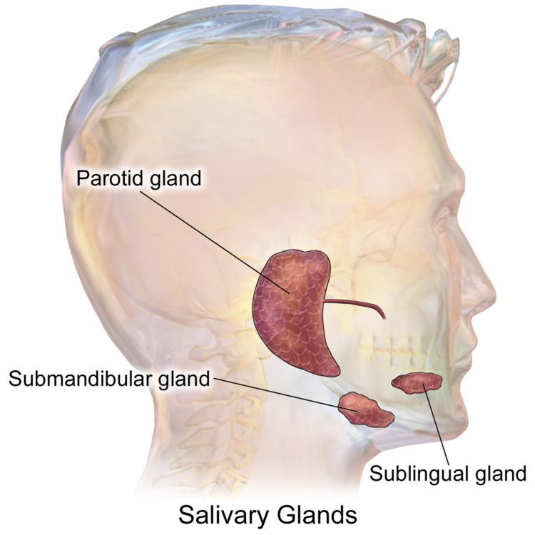
The salivary glands are detailed below:
Parotid glands
The two parotid glands are major salivary glands wrapped around the mandibular ramus in humans. The largest of the salivary glands, they secrete saliva to facilitate mastication and swallowing, and amylase to begin the digestion of starches. It is the serous type of gland which secretes the ptyalin. It enters the oral cavity via the parotid duct or Stensen duct. The glands are located posterior to the mandibular ramus and anterior to the mastoid process of the temporal bone. They are clinically relevant in dissections of facial nerve branches while exposing the different lobes of it since any iatrogenic lesion will result in either loss of action or strength of muscles involved in facial expression. They produce 20% of the total salivary content in the oral cavity.
Submandibular glands
The submandibular glands are a pair of major salivary glands located beneath the lower jaws, superior to the digastric muscles. The secretion produced is a mixture of both serous fluid and mucus, and enters the oral cavity via the submandibular duct or Wharton duct. Approximately 65-70% of saliva in the oral cavity is produced by the submandibular glands, even though they are much smaller than the parotid glands.You can usually feel this gland, as it is in the superficial cervical region and feels like a rounded ball. It is located about two fingers above the Adam's apple (laryngeal prominence) and about two inches apart under the chin.
Sublingual glands
The sublingual glands are a pair of major salivary glands located inferior to the tongue, anterior to the submandibular glands. The secretion produced is mainly mucous in nature; however, it is categorized as a mixed gland. Unlike the other two major glands, the ductal system of the sublingual glands does not have intercalated ducts and usually does not have striated ducts either, so saliva exits directly from 8-20 excretory ducts known as the Rivinus ducts. Approximately 5% of saliva entering the oral cavity comes from these glands.
Minor salivary glands
There are 800-1000 minor salivary glands located throughout the oral cavity within the submucosa of the oral mucosa in the tissue of the buccal, labial, and lingual mucosa, the soft palate, the lateral parts of the hard palate, and the floor of the mouth or between muscle fibers of the tongue. They are 1-2mm in diameter and unlike the major glands, they are not encapsulated by connective tissue, only surrounded by it. The gland has usually a number of acini connected in a tiny lobule. A minor salivary gland may have a common excretory duct with another gland, or may have its own excretory duct. Their secretion is mainly mucous in nature (except for Von Ebner glands- see next section) and have many functions such as coating the oral cavity with saliva. Problems with dentures are sometimes associated with minor salivary glands if there is dry mouth present (see further discussion). The minor salivary glands are innervated by the seventh cranial or facial nerve.
Von Ebner's glands
Von Ebner's glands are glands found in a trough circling the circumvallate papillae on the dorsal surface of the tongue near the terminal sulcus. They secrete a purely serous fluid that begins lipid hydrolysis. They also facilitate the perception of taste through secretion of digestive enzymes and proteins. The arrangement of these glands around the circumvallate papillae provides a continuous flow of fluid over great number of taste bud along the sides of the papillae. It is important for dissolving the food particles to be tested.
Nerve supply
Salivary glands are innervated, either directly or indirectly, by the parasympathetic and sympathetic arms of the autonomic nervous system. Parasympathetic stimulation evokes a copious flow of saliva. In contrast, sympathetic stimulation produces either a small flow, which is rich in protein, or no flow at all.
Microanatomy
The gland is internally divided into lobules. Blood vessels and nerves enter the glands at the hilum and gradually branch out into the lobules.
Acini
Secretory cells are found in a group, or acinus (plural, acini). Each acinus is located at the terminal part of the gland connected to the ductal system, with many acini within each lobule of the gland. Each acinus consists of a single layer of cuboidal epithelial cells surrounding a lumen, a central opening where the saliva is deposited after being produced by the secretory cells. The three forms of acini are classified in terms of the type of epithelial cell present and the secretory product being produced: serous, mucoserous and mucous.
Ducts
In the duct system, the lumina are formed by intercalated ducts, which in turn join to form striated ducts. These drain into ducts situated between the lobes of the gland (called interlobar ducts or secretory ducts). These are found on most major and minor glands (exception may be the sublingual gland).
All of the human salivary glands terminate in the mouth, where the saliva proceeds to aid in digestion. The saliva that salivary glands release is quickly inactivated in the stomach by the acid that is present there but the saliva also contains enzymes that are actually activated by the acid.
Clinical significance
A sialogram is a radiocontrast study of a salivary duct that may be used to investigate its function.
Salivary duct calculus may cause blockage of the ducts, causing pain and swelling of the gland because of cysts.
Saliva production may be pharmacologically stimulated by sialagogues (e.g., pilocarpin, cevimeline). It can also be suppressed by so-called antisialagogues (e.g., tricyclic antidepressants, SSRI, antihypertensives, polypharmacy). Many anti-cancer treatments may impair salivary flow such as chemotherapy and radiation therapy. Radiation therapy may cause permanent hyposalivation due to injury to the oral mucosa containing the salivary glands, resulting in dry mouth or xerostomia, whereas chemotherapy may cause only temporary salivary impairment.
Graft versus host disease after allogeneic bone marrow transplantation may manifest as dry mouth and many small mucoceles.
Tumours of the salivary glands may occur, including mucoepidermoid carcinoma.
Ageing of Salivary Glands
Aging of salivary glands show some structural changes:
In addition to that, there would be some changes in salivary contents:
However, there is no overall change in the amount of saliva secreted.
Other animals
The salivary glands of some species however, are modified to produce proteins; salivary amylase is found in many, but by no means all, bird and mammal species (including humans, as noted above). Furthermore, the venom glands of poisonous snakes, Gila monsters, and some shrews, are modified salivary glands. In other organisms such as insects, salivary glands are often used to produce biologically important proteins like silk or glues, and fly salivary glands contain polytene chromosomes that have been useful in genetic research.