
Carnegie stage 5b TE E5.7.1.0.0.0.4 | Days 9 MeSH sac A10.615.284.981 | |
 | ||
Latin vesicula umbilicalis; saccus vitellinus | ||
The yolk sac is a membranous sac attached to an embryo, formed by cells of the hypoblast adjacent to the embryonic disk. This is alternatively called the umbilical vesicle by the Terminologia Embryologica (TE), though yolk sac is far more widely used. The yolk sac is important in early embryonic blood supply, and much of it is incorporated into the primordial gut during the fourth week of development.
Contents
In humans
The yolk sac is the first element seen within the gestational sac during pregnancy, usually at 3 days gestation.
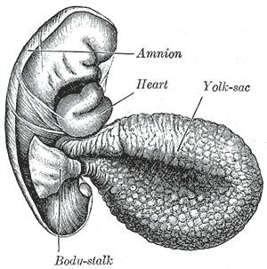
The yolk sac is situated on the ventral aspect of the embryo; it is lined by extra-embryonic endoderm, outside of which is a layer of extra-embryonic mesenchyme, derived from the mesoderm.
Blood is conveyed to the wall of the yolk sac by the primitive aorta, and after circulating through a wide-meshed capillary plexus, is returned by the vitelline veins to the tubular heart of the embryo. This constitutes the vitelline circulation, and by means of it nutritive material is absorbed from the yolk sac and conveyed to the embryo.
At the end of the fourth week the yolk sac presents the appearance of a small pear-shaped opening (traditionally called the umbilical vesicle), into the digestive tube by a long narrow tube, the vitelline duct.
The yolk sac can be seen in the afterbirth as a small, somewhat oval-shaped body whose diameter varies from 1 mm. to 5 mm.; it is situated between the amnion and the chorion and may lie on or at a varying distance from the placenta.
As a rule the duct undergoes complete obliteration during the seventh week, but in about two percent of cases its proximal part persists as a diverticulum from the small intestine, Meckel's diverticulum, which is situated about 60 cm proximal to the ileocecal valve, and may be attached by a fibrous cord to the abdominal wall at the umbilicus.
Sometimes a narrowing of the lumen of the ileum is seen opposite the site of attachment of the duct.
Histogenesis
The yolk sac starts forming during the second week of the embryonic development, at the same time as the shaping of the amniotic sac. The hypoblast starts proliferating laterally and descending. In the meantime Heuser's membrane, located on the opposite pole of the developing vesicle, starts its upward proliferation and meets the hypoblast.