
Latin Uterus | Greek Hystera | |
 | ||
Lymph Body and cervix to internal iliac lymph nodes, fundus to para-aortic lymph nodes, lumbar and superficial inguinal lymph nodes. | ||
The uterus (from Latin "uterus", plural uteri) or womb is a major female hormone-responsive reproductive sex organ of humans and most other mammals. In the human, the lower end of the uterus, the cervix, opens into the vagina, while the other end, the fundus, is connected to the fallopian tubes. It is within the uterus that the fetus develops during gestation. In the embryo the uterus develops from the paramesonephric ducts which fuse into the single organ known as a simplex uterus. The uterus has different forms in many other animals and in some it exists as two separate uteri known as a duplex uterus.
Contents
- Structure
- Regions
- Layers
- Support
- Major ligaments
- Axes
- Position
- Blood supply
- Nerve supply
- Development
- Function
- Clinical significance
- Uterus transplantation
- Other animals
- References
In English, the term uterus is used consistently within the medical and related professions, while the Germanic-derived term womb is also common in everyday contexts.
Structure
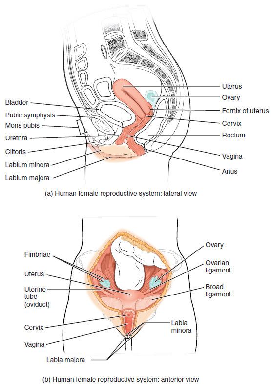
The uterus is located within the pelvic region immediately behind and almost overlying the bladder, and in front of the rectum. The human uterus is pear-shaped and about 7.6 cm (3 in.) long, 4.5 cm broad (side to side) and 3.0 cm thick. A typical adult uterus weighs about 60 grams. The uterus can be divided anatomically into four regions: The fundus, corpus (body), cervix and the internal os. The cervix protrudes into the vagina. The uterus is held in position within the pelvis by condensations of endopelvic fascia, which are called ligaments. These ligaments include the pubocervical, transverse cervical ligaments or cardinal ligaments, and the uterosacral ligaments. It is covered by a sheet-like fold of peritoneum, the broad ligament.
Regions
From outside to inside, the path to the uterus is as follows:
Layers
The three layers, from innermost to outermost, are as follows:
Support
The uterus is primarily supported by the pelvic diaphragm, perineal body and the urogenital diaphragm. Secondarily, it is supported by ligaments and the peritoneal ligament the broad ligament of uterus.
Major ligaments
It is held in place by several peritoneal ligaments, of which the following are the most important (there are two of each):
Axes
Normally the uterus lies in anteversion & anteflexion. In most women, the long axis of the uterus is bent forward on the long axis of the vagina, against the urinary bladder. This position is referred to as anteversion of the uterus. Furthermore, the long axis of the body of the uterus is bent forward at the level of the internal os with the long axis of the cervix. This position is termed anteflexion of the uterus. Uterus assumes anteverted position in 50% women, retroverted position in 25% women and rest have midposed uterus.
Position
The uterus is in the middle of the pelvic cavity in frontal plane (due to ligamentum latum uteri). The fundus does not surpass the linea terminalis, while the vaginal part of the cervix does not extend below interspinal line. The uterus is mobile and moves posteriorly under the pressure of a full bladder, or anteriorly under the pressure of a full rectum. If both are full, it moves upwards. Increased intraabdominal pressure pushes it downwards. The mobility is conferred to it by musculo-fibrous apparatus that consists of suspensory and sustentacular part. Under normal circumstances the suspensory part keeps the uterus in anteflexion and anteversion (in 90% of women) and keeps it "floating" in the pelvis. The meaning of these terms are described below:
Sustentacular part supports the pelvic organs and comprises the larger pelvic diaphragm in the back and the smaller urogenital diaphragm in the front.
The pathological changes of the position of the uterus are:
In cases where the uterus is "tipped", also known as retroverted uterus, women may have symptoms of pain during sexual intercourse, pelvic pain during menstruation, minor incontinence, urinary tract infections, fertility difficulties, and difficulty using tampons. A pelvic examination by a doctor can determine if a uterus is tipped.
Blood supply
The uterus is supplied by arterial blood both from the uterine artery and the ovarian artery. Another anastomotic branch may also supply the uterus from anastomosis of these two arteries.
Nerve supply
Afferent nerves supplying uterus are T11 and T12. Sympathetic supply is from hypogastric plexus and ovarian plexus. Parasympathetic supply is from second, third and fourth sacral nerves.
Development
Bilateral Müllerian ducts form during early fetal life. In males, MIF secreted from the testes leads to their regression. In females, these ducts give rise to the Fallopian tubes and the uterus. In humans the lower segments of the two ducts fuse to form a single uterus, however, in cases of uterine malformations this development may be disturbed. The different uterine forms in various mammals are due to various degrees of fusion of the two Müllerian ducts.
Function
The uterus is essential in sexual response by directing blood flow to the pelvis and ovaries, and to the external genitals, including the vagina, labia, and clitoris.
The reproductive function of the uterus is to accept a fertilized ovum which passes through the utero-tubal junction from the fallopian tube (uterine tube). The ovum divides to become a blastocyst, which implants into the endometrium, and derives nourishment from blood vessels which develop exclusively for this purpose. The fertilized ovum becomes an embryo, attaches to a wall of the uterus, creates a placenta, and develops into a fetus (gestates) until childbirth. Due to anatomical barriers such as the pelvis, the uterus is pushed partially into the abdomen due to its expansion during pregnancy. Even during pregnancy the mass of a human uterus amounts to only about a kilogram (2.2 pounds).
Clinical significance
A hysterectomy is the surgical removal of the uterus which may be carried out for a number of reasons including the ridding of tumours both benign and malignant. A complete hysterectomy involves the removal of the body, fundus, and cervix of the uterus. A partial hysterectomy may just involve the removal of the uterine body while leaving the cervix intact. It is the most commonly performed gynecological surgical procedure.
Some pathological states include:
Uterus transplantation
In 2012, the world's first womb transplant from a dead donor was performed on a Turkish woman who was born without a womb, but has her own ovaries. She is in good condition and the womb is functional. In the year 2000 in Saudi Arabia a similar transplant was performed, but from a live donor. Although womb transplants have been successful in animals such as mice, rats and sheep, the prevailing opinion in the field is that the risks are too great. Apart from risks of rejection of the new womb, there is concern that the drugs necessary for prevention of rejection of the donated womb might harm the fetus.
Other animals
Most animals that lay eggs, such as birds and reptiles, including most ovoviviparous species, have an oviduct instead of a uterus. However, recent research into the biology of the viviparous (not merely ovoviviparous) skink Trachylepis ivensi has revealed development of a very close analogue to eutherian mammalian placental development.
In monotremes, mammals which lay eggs, namely the platypus and the echidnas, either the term uterus or oviduct is used to describe the same organ, but the egg does not develop a placenta within the mother and thus does not receive further nourishment after formation and fertilization.
Marsupials have two uteri, each of which connect to a lateral vagina and which both use a third, middle "vagina" which functions as the birth canal. Marsupial embryos form a choriovitelline "placenta" (which can be thought of as something between a monotreme egg and a "true" placenta), in which the egg's yolk sac supplies a large part of the embryo's nutrition but also attaches to the uterine wall and takes nutrients from the mother's bloodstream.
The fetus usually develops fully in placental mammals and only partially in marsupials including kangaroos and opossums. In marsupials the uterus forms as a duplex organ of two uteri. In monotremes (egg-laying mammals) such as the platypus, the uterus is duplex and rather than nurturing the embryo, secretes the shell around the egg. It is essentially identical with the shell gland of birds and reptiles, with which the uterus is homologous.
In mammals, the four main forms of the uterus are: duplex, bipartite, bicornuate and simplex.
Two uteri usually form initially in a female and usually male fetus, and in placental mammals they may partially or completely fuse into a single uterus depending on the species. In many species with two uteri, only one is functional. Humans and other higher primates such as chimpanzees, usually have a single completely fused uterus, although in some individuals the uteri may not have completely fused.