
 | ||
A fetus is a stage in the prenatal development of viviparous organisms. In human development, a fetus or foetus (/ˈfiːtəs/; plural fetuses or foetuses) is a prenatal human between the embryonic state and birth. The fetal stage of development tends to be taken as beginning at the gestational age of eleven weeks, i.e. nine weeks after fertilization. In biological terms, however, prenatal development is a continuum, with no clear defining feature distinguishing an embryo from a fetus. The use of the term "fetus" generally implies that an embryo has developed to the point of being recognizable as a human; this is the point usually taken to be the ninth week after fertilization. A fetus is also characterized by the presence of all the major body organs, though they will not yet be fully developed and functional and some not yet situated in their final anatomical location.
Contents
Etymology
The word fetus (plural fetuses) is from the Latin fētus (“offspring”, “bringing forth”, “hatching of young”). The British, Irish, and Commonwealth spelling is foetus, which has been in use since at least 1594. It arose as a hypercorrection based on an incorrect etymology (i.e. due to insufficient knowledge of Latin) that may have originated with an error by Isidore of Seville in AD 620. This spelling is the most common in most Commonwealth nations, except in the medical literature, where fetus is used. The etymologically accurate original spelling fetus is used in Canada and the United States. In addition, fetus is now the standard English spelling throughout the world in medical journals. The spelling faetus was used historically.
Weeks 9 to 16 (2–1/4 to 4 months)
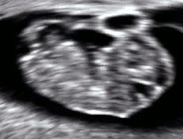
In humans, the fetal stage commences at the beginning of the ninth week. At the start of the fetal stage, the fetus is typically about 30 millimetres (1.2 in) in length from crown to rump, and weighs about 8 grams. The head makes up nearly half of the fetus' size. Breathing-like movement of the fetus is necessary for stimulation of lung development, rather than for obtaining oxygen. The heart, hands, feet, brain and other organs are present, but are only at the beginning of development and have minimal operation. The genitalia of the fetus starts to form and placenta becomes fully functional during week 9.
At this point in development, uncontrolled movements and twitches occur as muscles, the brain, and pathways begin to develop.
Weeks 17 to 25 (4–1/4 to 6–1/4 months)
A woman pregnant for the first time (primiparous), typically feels fetal movements at about 21 weeks, whereas a woman who has given birth at least once (multiparous), will typically feel movements by 20 weeks. By the end of the fifth month, the fetus is about 20 cm (8 inches) long.
Weeks 26 to 38 (6–1/2 to 9–1/2 months)
The amount of body fat rapidly increases. Lungs are not fully mature. Thalamic brain connections, which mediate sensory input, form. Bones are fully developed, but are still soft and pliable. Iron, calcium, and phosphorus become more abundant. Fingernails reach the end of the fingertips. The lanugo, or fine hair, begins to disappear, until it is gone except on the upper arms and shoulders. Small breast buds are present on both sexes. Head hair becomes coarse and thicker. Birth is imminent and occurs around the 38th week after fertilization. The fetus is considered full-term between weeks 36 and 40, when it is sufficiently developed for life outside the uterus. It may be 48 to 53 cm (19 to 21 inches) in length, when born. Control of movement is limited at birth, and purposeful voluntary movements develop all the way until puberty.
Variation in growth
There is much variation in the growth of the human fetus. When fetal size is less than expected, that condition is known as intrauterine growth restriction (IUGR) also called fetal growth restriction (FGR); factors affecting fetal growth can be maternal, placental, or fetal.
Maternal factors include maternal weight, body mass index, nutritional state, emotional stress, toxin exposure (including tobacco, alcohol, heroin, and other drugs which can also harm the fetus in other ways), and uterine blood flow.
Placental factors include size, microstructure (densities and architecture), umbilical blood flow, transporters and binding proteins, nutrient utilization and nutrient production.
Fetal factors include the fetus genome, nutrient production, and hormone output. Also, female fetuses tend to weigh less than males, at full term.
Fetal growth is often classified as follows: small for gestational age (SGA), appropriate for gestational age (AGA), and large for gestational age (LGA). SGA can result in low birth weight, although premature birth can also result in low birth weight. Low birth weight increases risk for perinatal mortality (death shortly after birth), asphyxia, hypothermia, polycythemia, hypocalcemia, immune dysfunction, neurologic abnormalities, and other long-term health problems. SGA may be associated with growth delay, or it may instead be associated with absolute stunting of growth.
Viability
Viability refers to a point in fetal development at which the fetus may survive outside the womb. The lower limit of viability is approximately 5-3/4 months gestational age and is usually later.
There is no sharp limit of development, age, or weight at which a fetus automatically becomes viable. According to data from 2003–05, survival rates are 20–35% for babies born at 23 weeks of gestation (5-3/4 months); 50–70% at 24-25 weeks (6 - 6-1/4 months); and >90% at 26-27 weeks (6-1/2 - 6-3/4 months) and over. It is rare for a baby weighing less than 1.1 pounds to survive.
When such premature babies are born, the main causes of perinatal mortality are that the respiratory system and the central nervous system are not completely differentiated. If given expert postnatal care, some fetuses weighing less than 1.1 pounds may survive, and are referred to as extremely low birth weight or immature infants. Preterm birth is the most common cause of perinatal mortality, causing almost 30 percent of neonatal deaths.
Fetal pain
Fetal pain, its existence and its implications are debated politically and academically. According to the conclusions of a review published in 2005, "Evidence regarding the capacity for fetal pain is limited but indicates that fetal perception of pain is unlikely before the third trimester." However, developmental neurobiologists argue that the establishment of thalamocortical connections (at about 6-1/2 months) is an essential event with regard to fetal perception of pain. Nevertheless, the perception of pain involves sensory, emotional and cognitive factors and it is "impossible to know" when pain is experienced, even if it is known when thalamocortical connections are established. Some authors argue that fetal pain is possible from the second half of pregnancy: “The available scientific evidence makes it possible, even probable, that fetal pain perception occurs well before late gestation” wrote KJS Anand in the journal of the IASP.
Whether a fetus has the ability to feel pain and suffering is part of the abortion debate. In the United States, for example, pro-life advocates have proposed legislation that would require providers of abortions to inform women that their fetuses may feel pain during the procedure and that would require each woman to accept or decline anesthesia for the fetus.
Circulatory system
The heart and blood vessels which form the circulatory system, form relatively early during embryonic development, but continue to grow and develop in complexity in the growing fetus. A functional circulatory system is a biological necessity, since mammalian tissues can not grow more than a few cell layers thick without an active blood supply. The prenatal circulation of blood is different than the postnatal circulation, mainly because the lungs are not in use. The fetus obtains oxygen and nutrients from the mother through the placenta and the umbilical cord.
Blood from the placenta is carried to the fetus by the umbilical vein. About half of this enters the fetal ductus venosus and is carried to the inferior vena cava, while the other half enters the liver proper from the inferior border of the liver. The branch of the umbilical vein that supplies the right lobe of the liver first joins with the portal vein. The blood then moves to the right atrium of the heart. In the fetus, there is an opening between the right and left atrium (the foramen ovale), and most of the blood flows from the right into the left atrium, thus bypassing pulmonary circulation. The majority of blood flow is into the left ventricle from where it is pumped through the aorta into the body. Some of the blood moves from the aorta through the internal iliac arteries to the umbilical arteries, and re-enters the placenta, where carbon dioxide and other waste products from the fetus are taken up and enter the woman's circulation.
Some of the blood from the right atrium does not enter the left atrium, but enters the right ventricle and is pumped into the pulmonary artery. In the fetus, there is a special connection between the pulmonary artery and the aorta, called the ductus arteriosus, which directs most of this blood away from the lungs (which aren't being used for respiration at this point as the fetus is suspended in amniotic fluid).
Postnatal development
With the first breath after birth, the system changes suddenly. Pulmonary resistance is reduced dramatically, prompting more blood to move into the pulmonary arteries from the right atrium and ventricle of the heart and less to flow through the foramen ovale into the left atrium. The blood from the lungs travels through the pulmonary veins to the left atrium, producing an increase in pressure that pushes the septum primum against the septum secundum, closing the foramen ovale and completing the separation of the newborn's circulatory system into the standard left and right sides. Thereafter, the foramen ovale is known as the fossa ovalis.
The ductus arteriosus normally closes within one or two days of birth, leaving the ligamentum arteriosum, while the umbilical vein and ductus venosus usually closes within two to five days after birth, leaving, respectively, the liver's ligamentum teres and ligamentum venosus.
Differences between fetal and postnatal
Remnants of the fetal circulation can be found in the adult.
In addition to differences in circulation, the developing fetus also employs a different type of oxygen transport molecule in its hemoglobin from that when it is born and breathing its own oxygen. Fetal hemoglobin enhances the fetus' ability to draw oxygen from the placenta. Its oxygen-hemoglobin dissociation curve is shifted to the left, meaning that it is able to absorb oxygen at lower concentrations than adult hemoglobin. This enables fetal hemoglobin to absorb oxygen from adult hemoglobin in the placenta, where the oxygen pressure is lower than at the lungs. Until around six months' old, the human infant's hemoglobin molecule is made up of two alpha and two gamma chains (2α2γ). The gamma chains are gradually replaced by beta chains until the molecule becomes hemoglobin A with its two alpha and two beta chains (2α2β).
Immune system
The placenta functions as a maternal-fetal barrier against the transmission of microbes. When this is insufficient, mother-to-child transmission of infectious diseases can occur.
Maternal IgG antibodies cross the placenta, giving the fetus passive immunity against those diseases for which the mother has antibodies. This transfer of antibodies in humans begins as early as the fifth month (gestational age) and certainly by the sixth month.
Developmental problems
A developing fetus is highly susceptible to anomalies in its growth and metabolism, increasing the risk of birth defects. One area of concern is the pregnant woman's lifestyle choices made during pregnancy. Diet is especially important in the early stages of development. Studies show that supplementation of the woman's diet with folic acid reduces the risk of spina bifida and other neural tube defects. Another dietary concern is whether breakfast is eaten. Skipping breakfast could lead to extended periods of lower than normal nutrients in the woman's blood, leading to a higher risk of prematurity, or other birth defects in the fetus. During this time alcohol consumption may increase the risk of the development of fetal alcohol syndrome, a condition leading to intellectual disability in some infants.
Smoking during pregnancy may also lead to reduced birth weight. Low birth weight is defined as 2500 grams (5.5 lb). Low birth weight is a concern for medical providers due to the tendency of these infants, described as "premature by weight", to have a higher risk of secondary medical problems. Some research shows that fetal ultrasounds (including Doppler, 3D/4D ultrasound and 2D ultrasound) can have negative effect on birth weight and neurodevelopment.
Congenital anomalies are acquired before birth. Infants with certain congenital anomalies of the heart can survive only as long as the ductus remains open: in such cases the closure of the ductus can be delayed by the administration of prostaglandins to permit sufficient time for the surgical correction of the anomalies. Conversely, in cases of patent ductus arteriosus, where the ductus does not properly close, drugs that inhibit prostaglandin synthesis can be used to encourage its closure, so that surgery can be avoided.
Legal issues
Abortion of a human pregnancy is legal and/or tolerated in most countries, although with gestational time limits that normally prohibit late-term abortions.
Other animals
A fetus is a stage in the prenatal development of viviparous organisms. This stage lies between embryogenesis and birth. Many vertebrates have fetal stages, ranging from most mammals to many fish. In addition, some invertebrates bear live young, including some species of onychophoran and many arthropods. The prevalence of convergent evolution to the fetal stage shows that it is relatively easy to develop. Indeed, it presumably originates from a delay of egg release, with the eggs being hatched inside the parent before being laid. Over time, the robustness of the egg wall can be decreased until it becomes little more than a sac.
The fetuses of most mammals are situated similarly to the human fetus within their mothers. However, the anatomy of the area surrounding a fetus is different in litter-bearing animals compared to humans: each fetus of a litter-bearing animal is surrounded by placental tissue and is lodged along one of two long uteri instead of the single uterus found in a human female.
Development at birth varies considerably among animals, and even among mammals. Altricial species are relatively helpless at birth and require considerable parental care and protection. In contrast, precocial animals are born with open eyes, have hair or down, have large brains, and are immediately mobile and somewhat able to flee from, or defend themselves against, predators. Primates are precocial at birth, with the exception of humans.
The duration of gestation in placental mammals (i.e. mammals other than monotremes and marsupials) varies from 18 days in jumping mice to 23 months in elephants. Generally speaking, fetuses of larger land mammals require longer gestation periods.
The benefits of a fetal stage means that young are more developed when they are born. Therefore, they may need less parental care and may be better able to fend for themselves. However, carrying fetuses exerts costs on the mother, who must take on extra food to fuel the growth of her offspring, and whose mobility and comfort may be affected (especially toward the end of the fetal stage).
In some instances, the presence of a fetal stage may allow organisms to time the birth of their offspring to a favorable season.