
Latin vesica urinaria | ||
 | ||
Similar Cystitis glandularis, Urinary bladder disease, Inferior vesical artery | ||
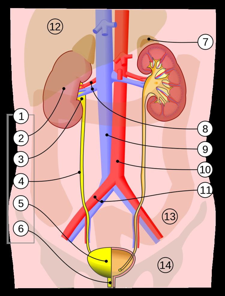
The urinary bladder is a hollow muscular organ that collects urine from the kidneys before disposal by urination. A hollow muscular, and distensible (or elastic) organ, the bladder sits on the pelvic floor. Urine enters the bladder via the ureters and exits via the urethra. The typical human bladder capacity is between 300 and 500 mL (10.14 and 16.91 fl oz).
Contents
Structure
The bladder is a hollow muscular organ situated at the base of the pelvis. Urine collects in the bladder from the two ureters, which open into the bladder at its back and connect to the kidneys. Urine leaves the bladder via the urethra, a single muscular tube which ends in the urethral orifice. Anatomically, the bladder is divided into a fundus at the top, two ureteric orifices, and an opening for the urethra which surrounds the trigone of the bladder. In men, the prostate gland lies outside the opening for the urethra.
The bladder is situated below the peritoneal cavity near the pelvic floor. In men, it lies in front of the rectum, separated by a space. In women, it lies in front of the uterus.
Histology
The outside of the bladder is protected by a serous membrane. The bladder wall itself is smooth muscle. The inner side of the bladder is lined with a mucosal membrane consisting of a surface glycocalyx that protects the cells beneath it from urine, the urothelium (a form of transitional epithelium), a basement membrane, and the sub-urothelium.
Detrusor muscle
The detrusor muscle is a layer of the urinary bladder wall made of smooth muscle fibers arranged in spiral, longitudinal, and circular bundles. When the bladder is stretched, this signals the parasympathetic nervous system to contract the detrusor muscle. This encourages the bladder to expel urine through the urethra. A meta-analysis on the effect of voiding position on urodynamics in males found that sitting down allows for improved contraction of the detrusor muscle.
Lymphatic supply
The fundus of the bladder is lymphatically drained by the external iliac lymph nodes.
Nerve supply
The bladder receives motor innervation from both sympathetic fibers, most of which arise from the hypogastric plexuses and nerves, and parasympathetic fibers, which come from the pelvic splanchnic nerves and the inferior hypogastric plexus.
Sensation from the bladder is transmitted to the central nervous system (CNS) via general visceral afferent fibers (GVA). GVA fibers on the superior surface follow the course of the sympathetic efferent nerves back to the CNS, while GVA fibers on the inferior portion of the bladder follow the course of the parasympathetic efferents.
For the urine to exit the bladder, both the autonomically controlled internal sphincter and the voluntarily controlled external sphincter must be opened. Problems with these muscles can lead to incontinence.
Development
The human urinary bladder is derived from the urogenital sinus, and it is initially continuous with the allantois. In males, the base of the bladder lies between the rectum and the pubic symphysis. It is superior to the prostate, and separated from the rectum by the rectovesical excavation. In females, the bladder sits inferior to the uterus and anterior to the vagina; thus, its maximum capacity is lower than in males. It is separated from the uterus by the vesicouterine excavation. In infants and young children, the urinary bladder is in the abdomen even when empty.
Function
Urine, excreted by the kidneys, collects in the bladder before disposal by urination. The urinary bladder usually holds 300-350 ml of urine. As urine accumulates, the rugae flatten and the wall of the bladder thins as it stretches, allowing the bladder to store larger amounts of urine without a significant rise in internal pressure.
Clinical significance
Frequent urination can be due to excessive urine production, small bladder capacity, irritability or incomplete emptying. Males with an enlarged prostate urinate more frequently. One definition of overactive bladder is when a person urinates more than eight times per day, though there can be other causes of urination frequency. Though both urinary frequency and volumes have been shown to have a circadian rhythm, meaning day and night cycles, it is not entirely clear how these are disturbed in the overactive bladder.
Disorders of or related to the bladder include:
Other animals
Bladders occur throughout much of the animal kingdom, but are very diverse in form, such as the swim bladder in fish, and in some cases are not homologous with the urinary bladder in humans. The urinary bladder of chelonians is very thin and cystoscopy permits visualisation of internal organs. The pig bladder is very similar to the human bladder.