
Latin Peritoneum Code TH H3.04.08.0.00001 TA A10.1.02.002 | MeSH A01.047.025.600 Dorlands/Elsevier Peritoneum FMA 9584 | |
 | ||
The peritoneum /ˌpɛrᵻtəˈniːəm/ is the serous membrane that forms the lining of the abdominal cavity or coelom in amniotes and some invertebrates, such as annelids. It covers most of the intra-abdominal (or coelomic) organs, and is composed of a layer of mesothelium supported by a thin layer of connective tissue. This peritoneal lining of the cavity supports many of the abdominal organs and serves as a conduit for their blood vessels, lymphatic vessels, and nerves.
Contents
- Types
- Subdivisions
- Other ligaments and folds
- Classification of abdominal structures
- Development
- Peritoneal dialysis
- Peritonitis
- Primary peritoneal carcinoma
- Etymology
- References
The abdominal cavity (the space bounded by the vertebrae, abdominal muscles, diaphragm, and pelvic floor) should not be confused with the intraperitoneal space (located within the abdominal cavity, but wrapped in peritoneum). The structures within the intraperitoneal space are called "intraperitoneal" (e.g., the stomach and intestines), the structures in the abdominal cavity that are located behind the intraperitoneal space are called "retroperitoneal" (e.g., the kidneys), and those structures below the intraperitoneal space are called "subperitoneal" or "infraperitoneal" (e.g., the bladder).
Types
Although they ultimately form one continuous sheet, two layers of peritoneum and a potential space between them are referenced:
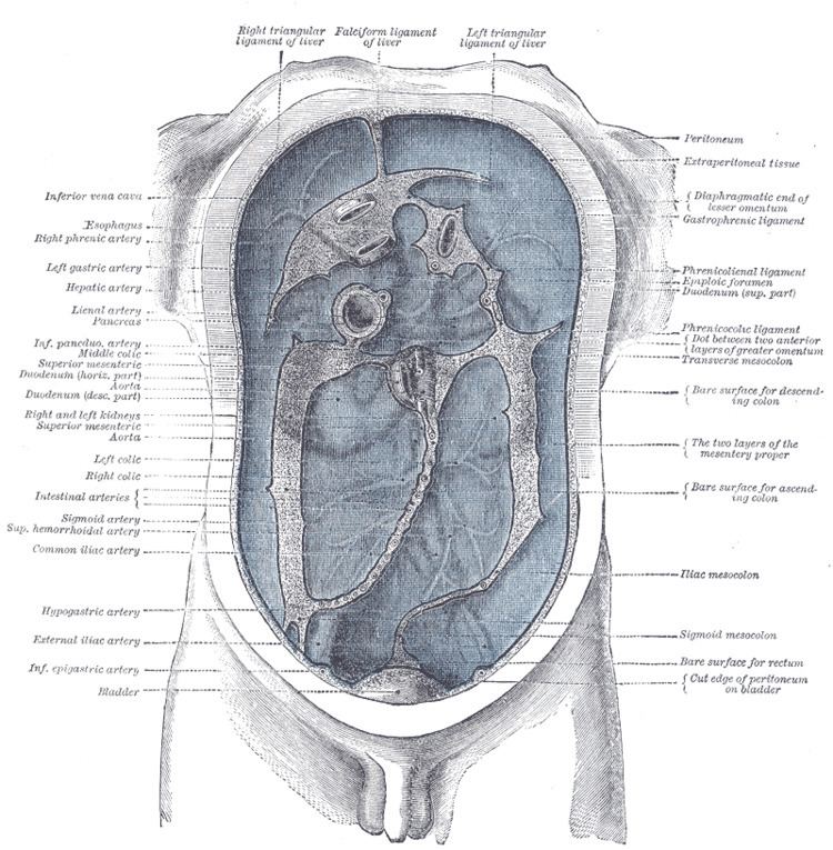
Subdivisions
Peritoneal folds are omenta, mesenteries and ligaments; they connect organs to each other or to the abdominal wall. There are two main regions of the peritoneal cavity, connected by the epiploic foramen (also known as the omental foramen or foramen of winslow):
The mesentery is the part of the peritoneum through which most abdominal organs are attached to the abdominal wall and supplied with blood and lymph vessels and nerves.
Other ligaments and folds
In addition, in the pelvic cavity there are several structures that are usually named not for the peritoneum, but for the areas defined by the peritoneal folds:
Classification of abdominal structures
The structures in the abdomen are classified as intraperitoneal, retroperitoneal or infraperitoneal depending on whether they are covered with visceral peritoneum and whether they are attached by mesenteries (mensentery, mesocolon).
Structures that are intraperitoneal are generally mobile, while those that are retroperitoneal are relatively fixed in their location.
Some structures, such as the kidneys, are "primarily retroperitoneal", while others such as the majority of the duodenum, are "secondarily retroperitoneal", meaning that structure developed intraperitoneally but lost its mesentery and thus became retroperitoneal.
Development
The peritoneum develops ultimately from the mesoderm of the trilaminar embryo. As the mesoderm differentiates, one region known as the lateral plate mesoderm splits to form two layers separated by an intraembryonic coelom. These two layers develop later into the visceral and parietal layers found in all serous cavities, including the peritoneum.
As an embryo develops, the various abdominal organs grow into the abdominal cavity from structures in the abdominal wall. In this process they become enveloped in a layer of peritoneum. The growing organs "take their blood vessels with them" from the abdominal wall, and these blood vessels become covered by peritoneum, forming a mesentery.
Peritoneal folds develop from the ventral and dorsal mesentery of the embryo.
Peritoneal dialysis
In one form of dialysis, called peritoneal dialysis, a glucose solution is sent through a tube into the peritoneal cavity. The fluid is left there for a prescribed amount of time to absorb waste products, and then removed through the tube. The reason for this effect is the high number of arteries and veins in the peritoneal cavity. Through the mechanism of diffusion, waste products are removed from the blood.
Peritonitis
Peritonitis is the inflammation of the peritoneum. It is more commonly associated to infection from a punctured organ of the abdominal cavity. It can also be provoked by the presence of fluids that produce chemical irritation, such as gastric acid or pancreatic juice. Peritonitis causes fever, tenderness, and pain in the abdominal area, which can be localized or diffuse. The treatment involves rehydration, administration of antibiotics, and surgical correction of the underlying cause. Mortality is higher in the elderly and if present for a prolonged time.
Primary peritoneal carcinoma
Primary peritoneal cancer is a cancer of the cells lining the peritoneum.
Etymology
Peritoneum is derived from Greek via Latin. Peri- means around, while -ton- refers to stretching. Thus, peritoneum means stretched around or stretched over.