
Specialty pulmonology | ||
 | ||
Synonyms pleuritis, pleuritic chest pain Medication paracetamol (acetaminophen), ibuprofen | ||
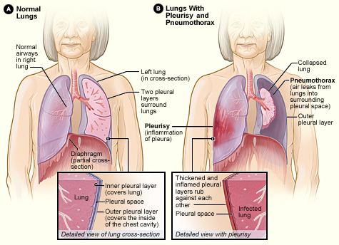
Pleurisy, also known as pleuritis, is inflammation of the membranes (pleurae) that surround the lungs and line the chest cavity. This can result in a sharp chest pain with breathing. Occasionally the pain may be a constant dull ache. Other symptoms may include shortness of breath, cough, fever, or weight loss depending on the underlying cause.
Contents
- Signs and symptoms
- Causes
- Diagnosis
- Physical examination
- Diagnostic tests
- Chest x ray
- Blood test
- ECG
- Ultrasound
- Computed tomography CT scan
- Magnetic resonance imaging MRI
- Arterial blood gas
- Thoracentesis
- Biopsy
- Treatment
- Procedures
- Medications
- Lifestyle changes
- Treating the cause
- Alternative treatments
- Related problems
- Pleural effusion
- Pneumothorax
- Hemothorax
- Prognosis
- Notable cases
- References
The most common cause is a viral infection. Other causes include pneumonia, pulmonary embolism, autoimmune disorders, lung cancer following heart surgery, pancreatitis, chest trauma, and asbestosis. Occasionally the cause remains unknown. The underlying mechanism involves the rubbing together of the pleurae instead of smooth gliding. Other conditions that can produce similar symptoms include pericarditis, heart attack, cholecystitis, and pneumothorax. Diagnosis may include a chest X-ray, electrocardiogram (ECG), and blood tests.
Treatment depends on the underlying cause. Paracetamol and ibuprofen may be used to help with the pain. Incentive spirometry may be recommended to encourage larger breaths. About one million people are affected in the United States each year. Descriptions of the condition date from at least as early as 400 BC by Hippocrates.
Signs and symptoms
The defining symptom of pleurisy is a sudden sharp, stabbing, burning or dull pain in the right or left side of the chest during breathing, especially when one inhales and exhales. It feels worse with deep breathing, coughing, sneezing, or laughing. The pain may stay in one place, or it may spread to the shoulder or back. Sometimes, it becomes a fairly constant dull ache.
Depending on its cause, pleuritic chest pain may be accompanied by other symptoms:
Causes
The pleural space can be invaded by fluid, air, and particles from different parts of the body which fairly complicates the diagnosis. Viral infection (coxsackie B virus, HRSV, CMV, adenovirus, EBV, parainfluenza, influenza) is the most common cause of pleurisy. However, many other different conditions can cause pleuritic chest pain:
When the space between the pleurae starts to fill with fluid, as in pleural effusion, the chest pain can be eased but a shortness of breath can result, since the lungs need room to expand during breathing. Some cases of pleuritic chest pain are idiopathic, which means that the exact cause cannot be determined.
Diagnosis
A diagnosis of pleurisy or another pleural condition is based on a medical history, physical examinations, and diagnostic tests. The goals are to rule out other sources of the symptoms and to find the cause of the pleurisy so that the underlying disorder can be treated.
Physical examination
A doctor uses a stethoscope to listen to the breathing. This method detects any unusual sounds in the lungs. A person with pleurisy may have inflamed layers of the pleurae that make a rough, scratchy sound as they rub against each other during breathing. This is called pleural friction rub.
Diagnostic tests
Depending on the results of the physical examination, diagnostic tests are sometimes performed.
Chest x-ray
A chest x-ray takes a picture of the heart and lungs. It may show air or fluid in the pleural space. It also may show the cause (e.g., pneumonia, a fractured rib, or a lung tumor) of the pleurisy.
Sometimes an x-ray is taken while lying on the painful side. This may show fluid, as well as changes in fluid position, that did not appear in the vertical x-ray.
Blood test
Blood tests can detect bacterial or viral infections, pneumonia, rheumatic fever, a pulmonary embolism, or lupus.
ECG
Electrocardiography test can determine if a heart condition contributes to the symptoms.
Ultrasound
Ultrasonography uses sound waves to create an image. It may show where fluid is located in the chest. It also can show some tumors. Although ultrasound may detect fluid around the lungs, also known as a pleural effusion, sound waves are scattered by air. Therefore, an actual picture of the lungs cannot be obtained with ultrasonography.
Computed tomography (CT) scan
A CT scan provides a computer-generated picture of the lungs that can show pockets of fluid. It also may show signs of pneumonia, a lung abscess, or a tumor.
Magnetic resonance imaging (MRI)
Magnetic resonance imaging (MRI), also called nuclear magnetic resonance (NMR) scanning, uses powerful magnets to show pleural effusions and tumors.
Arterial blood gas
In arterial blood-gas sampling, a small amount of blood is taken from an artery, usually in the wrist. The blood is then checked for oxygen and carbon-dioxide levels. This test shows how well the lungs are taking in oxygen.
Thoracentesis
Once the presence of an excess fluid in the pleural cavity, or pleural effusion, is suspected and location of fluid is confirmed, a sample of fluid can be removed for testing. The procedure to remove fluid in the chest is called a diagnostic thoracentesis. The doctor inserts a small needle or a thin, hollow, plastic tube in the chest wall and withdraws fluid.
Thoracentesis can be done in the doctor's office or at the hospital. Ultrasound is used to guide the needle to the fluid that is trapped in small pockets around the lungs.
Thoracentesis usually does not cause serious complications. Generally, a chest x-ray is done after the procedure to evaluate the lungs. Possible complications of thoracentesis include the following:
The lung fluid is examined under a microscope and is evaluated for the presence of chemicals and for its color and texture. The degree of clarity is an indicator of infection, cancer, or other conditions that may be causing the buildup of fluid or blood in the pleural space.
Biopsy
If tuberculosis or cancer is suspected, a small piece of the pleura may be examined under a microscope to make a definitive diagnosis. This is called a biopsy.
Several approaches to taking tissue samples are available
- Insertion of a needle through the skin on the chest to remove a small sample of the outer layer of the pleura.
- Insertion of a small tube with a light on the end (endoscope) into tiny cuts in the chest wall in order to visualize the pleura. Small pieces of tissue can be biopsied though the endoscope.
- Removal of a sample of the pleura through a small cut in the chest wall. This is called an open pleural biopsy. It is usually done if the sample from the needle biopsy is too small for an accurate diagnosis.
Treatment
Treatment has several goals:
Procedures
If large amounts of fluid, air, or blood are not removed from the pleural space, they may cause the lung to collapse.
The surgical procedures used to drain fluid, air, or blood from the pleural space are as follows:
Medications
A couple of medications are used to relieve pleurisy symptoms:
There may be a role for the use of corticosteroids (for tuberculous pleurisy), tacrolimus (Prograf) and methotrexate (Trexall, Rheumatrex) in the treatment of pleurisy. Further studies are needed.
Lifestyle changes
The following may be helpful in the management of pleurisy:
Treating the cause
Ideally, the treatment of pleurisy is aimed at eliminating the underlying cause of the disease.
The treatment for pleurisy depends on its origin and is prescribed by a physician on a base of an individual assessment. Paracetamol (acetaminophen) and amoxicillin, or other antibiotics in case of bacterial infections, are common remedies dispensed by doctors to relieve the initial symptoms and pain in the chest, while viral infections are self-limited. Non-steroidal anti-inflammatory drugs (NSAIDs), preferably indometacin, are usually employed as pain control agents.
Alternative treatments
A number of alternative or complementary medicines are being investigated for their anti-inflammatory properties, and their use in pleurisy. At this time, clinical trials of these compounds have not been performed.
Extracts from the Brazilian folk remedy Wilbrandia ebracteata ("Taiuia") have been shown to reduce inflammation in the pleural cavity of mice. The extract is thought to inhibit the same enzyme, cyclooxygenase-2 (COX-2), as the non-steroidal anti-inflammatory drugs.
Related problems
Pleurisy is often associated with complications that affect the pleural space.
Pleural effusion
In some cases of pleurisy, excess fluid builds up in the pleural space. This is called a pleural effusion. The buildup of fluid usually forces the two layers of the pleura apart so they don't rub against each other when breathing. This can relieve the pain of pleurisy. A large amount of extra fluid can push the pleura against the lung until the lung, or a part of it, collapses. This can make it hard to breathe.
In some cases of pleural effusion, the extra fluid gets infected and turns into an abscess. This is called an empyema.
Pleural effusion involving fibrinous exudates in the fluid may be called fibrinous pleurisy. It sometimes occurs as a later stage of pleurisy.
A person can develop a pleural effusion in the absence of pleurisy. For example, pneumonia, heart failure, cancer, or a pulmonary embolism can lead to a pleural effusion.
Pneumothorax
Air or gas also can build up in the pleural space. This is called a pneumothorax. It can result from acute lung injury or a lung disease like emphysema. Lung procedures, like surgery, drainage of fluid with a needle, examination of the lung from the inside with a light and a camera, or mechanical ventilation, also can cause a pneumothorax.
The most common symptom is sudden pain in one side of the lung and shortness of breath. A pneumothorax also can put pressure on the lung and cause it to collapse.
If the pneumothorax is small, it may go away on its own. If large, a chest tube is placed through the skin and chest wall into the pleural space to remove the air.
Hemothorax
Blood also can collect in the pleural space. This is called hemothorax. The most common cause is injury to the chest from blunt force or surgery on the heart or chest. Hemothorax also can occur in people with lung or pleural cancer.
Hemothorax can put pressure on the lung and force it to collapse. It also can cause shock, a state of hypoperfusion in which an insufficient amount of blood is able to reach the organs.
Prognosis
Pleurisy and other disorders of the pleurae can be serious, depending on what caused them. Generally, pleurisy treatment has an excellent prognosis, but if left untreated it can cause severe complications. For example, a resulting pulmonary heart disease cor pulmonale, which manifests itself with an inflammation of the arms and legs, can lead to heart failure. If the conditions that caused the pleurisy or other pleural disorders were adequately diagnosed and treated early, one can expect a full recovery. Help of a pulmonologist (respiratory physician in the U.K. and Australia) may be enlisted to address the underlying cause and chart post-illness rehabilitation.