
Specialty emergency medicine ICD-9-CM 511.8, 860 MedlinePlus 000126 | ICD-10 J94.2, S27.1 DiseasesDB 19762 eMedicine med/2915 ped/971 | |
 | ||
A hemothorax (hemo- + thorax) (or haemothorax or haemorrhagic pleural effusion) is a type of pleural effusion in which blood accumulates in the pleural cavity. This excess fluid can interfere with normal breathing by limiting the expansion of the lungs.
Contents
Signs and symptoms
Hemothorax tends to occur following blunt or penetrating trauma to the thorax or thoracoabdominal area. It may also follow thoracic surgery, or may be spontaneous. Chest pain, dyspnea, and tachypnea are common presenting features. Other symptoms of hemothorax are dependent on the mechanism of injury, but may include:
Cause and presentation
Its cause is usually traumatic, from a blunt or penetrating injury to the thorax, resulting in a rupture of the serous membrane either lining the thorax or covering the lungs. This rupture allows blood to spill into the pleural space, equalizing the pressures between it and the lungs. Blood loss may be massive in people with these conditions, as each side of the thorax can hold 30 to 40% of a person's blood volume or 1.5 to 2 L per side in the average adult. Even minor injury to the chest wall can lead to significant hemothorax.
Less frequently, hemothorax occurs spontaneously. A major vascular cause of hemothorax is aortic dissection or rupture of thoracic aortic aneurysms. It may also follow surgical intervention in the thoracic area. Infrequently, patients with pneumothorax may develop spontaneous hemothorax. Spontaneous hemothorax or hemopneumothorax may be occur with endometriosis, if endometrial tissue implants on the pleural surface, then bleeds in response to cyclical hormonal changes in menstruating women.
Mechanism
The thoracic cavity is a chamber within the chest, containing the lungs, heart, and numerous major blood vessels. The parietal pleura lines the chest wall, while the visceral pleura covers the outside of the lungs. The visceral and parietal pleura are separated by a thin layer serous fluid. When a hemothorax occurs, blood enters the pleural cavity, which normally only contains a small amount of pleural fluid. As blood builds up within the pleural cavity, it begins to interfere with the normal movement of the lungs, resulting in dypsnea, poor ventilation, and abnormal oxygenation.
As a result of the body's response to the presence of blood in the pleura, a small hemothorax may lead to a bloody pleural effusion. When enzymes in the pleural fluid begin to break down blood clots, the protein concentration of the pleural fluid increases, and as a result the osmotic pressure of the pleural cavity increases, causing fluid to flow into the pleural cavity from the surrounding tissues. The resulting increase in fluid volume in the pleural cavity interferes with normal breathing and oxygenation, resulting in dypsnea and tachypnea.
Diagnosis
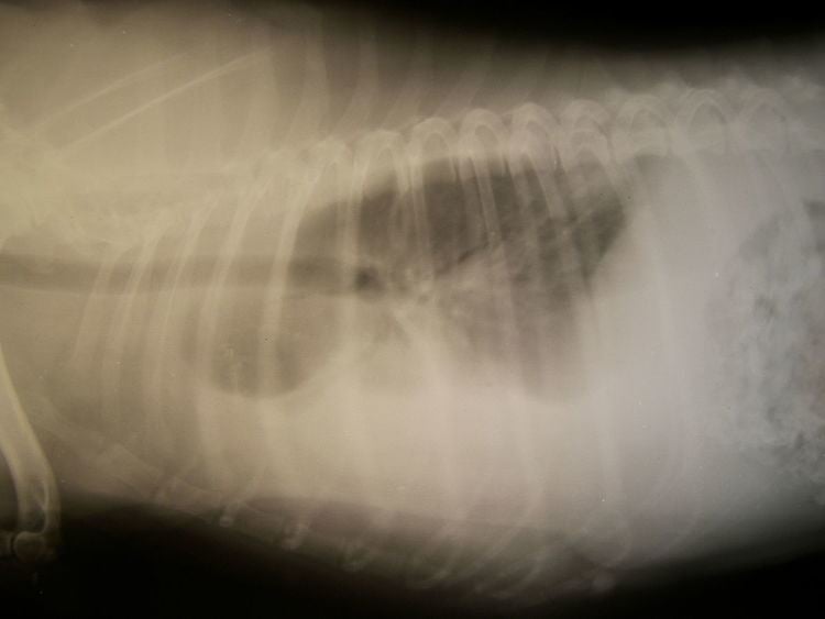
Chest radiography is the preferred means of initial diagnosis for hemothorax. Upright radiography is preferred but supine films may be taken when upright radiography is not feasible due to the clinical situation. Tube thoracostomy may be done prior to imaging when patients have sustained blunt or penetrating thoracic trauma and display unstable hemodynamics, have respiratory failure with absent or decreased breath sounds, show tracheal deviation, or have serious penetrating injuries. In upright radiography, hemothorax is suggested by blunting of the costophrenic angle or partial or complete opacification of the hemithorax, in which the lateral side of the chest appears bright and the lung appears pushed away toward the center; the air-filled lung normally appears as a dark space on radiographic film. In the case of a small hemothorax, several hundred milliliters of blood can be hidden by the diaphragm and abdominal viscera. In supine patients, signs of hemothorax may also be subtle on radiographic film, because the blood will layer in the pleural space, and can be seen as a haziness in one half of the thorax relative to the other side.
Ultrasonography is also used for detection of hemothorax and other pleural effusions, particularly in the critical care and trauma settings, because it provides rapid, reliable results in order to make a diagnosis in an emergency situation. Computed tomography (CT or CAT) scans can detect much smaller amounts of fluid than chest radiography, but computed tomography is not a primary method of diagnosis with in the trauma setting, due to the time required for imaging, the requirement that a patient remain supine, and the need to transport a critically ill patient to the scanner.
Prognosis
If left untreated, the condition can progress to a point where the blood accumulation begins to put pressure on the mediastinum and the trachea, effectively limiting the amount that the heart's ventricles are able to fill. The condition can cause the trachea to deviate, or move, toward the unaffected side.
Management
A hemothorax is managed by removing the source of bleeding and by draining the blood already in the thoracic cavity. Blood in the cavity can be removed by inserting a drain (chest tube) in a procedure called a tube thoracostomy. Generally, the thoracostomy tube is placed between the ribs in the sixth or seventh intercostal space at the mid-axillary line. Usually the lung will expand and the bleeding will stop after a chest tube is inserted.
The blood in the chest can thicken as the clotting cascade is activated when the blood leaves the blood vessels and comes into contact with the pleural surface, injured lung or chest wall, or with the chest tube. As the blood thickens, it can clot in the pleural space (leading to a retained hemothorax) or within the chest tube, leading to chest tube clogging or occlusion. Chest tube clogging or occlusion can lead to worse outcomes as it prevents adequate drainage of the pleural space, contributing to the problem of retained hemothorax. In this case, patients can be hypoxic, short of breath, or in some cases, the retained hemothorax can become infected (empyema).
Retained hemothorax occurs when blood remains in the pleural space, and is a risk factor for the development of complications, including the accumulation of pus in the pleural space and fibrothorax. It is treated by inserting a second chest tube or by drainage by video-assisted thoracoscopy. Fibrolytic therapy has also been studied as a treatment.
When hemothorax is treated with a chest tube, it is important that it maintain its function so that the blood cannot clot in the chest or the tube. If clogging occurs, internal chest tube clearing can be performed using an open or closed technique. Manual manipulation, which may also be called milking, stripping, or tapping, of chest tubes is commonly performed to maintain an open tube, but no conclusive evidence has demonstrated that any of these techniques are more effective than the others, or that they improve chest tube drainage. When chest tube clogging does occur, it can result in retained blood syndrome, especially after heart surgery and thoracic surgery.
In some cases bleeding continues and surgery is necessary to stop the source of bleeding. For example, if the hemothorax was caused by aortic rupture in high energy trauma, surgical intervention is mandatory.