
Group Group V ((−)ssRNA) Rank Family | Scientific name Paramyxoviridae | |
 | ||
Lower classifications Measles virus, Sendai virus, Avian pneumovirus, Henipavirus, Mumps virus | ||
Micro vet 2015 1 aula 7 fam lia paramyxoviridae
Paramyxoviridae (from Greek para-, beyond, -myxo-, mucus or slime, plus virus, from Latin poison, slime, thus meaning "slime beyond slime"; sometimes abbreviated PMV) is a family of viruses in the order Mononegavirales. Humans, vertebrates, and birds serve as natural hosts. There are currently 38 species in this family, divided among 7 genera. Diseases associated with this negative-sense single-stranded RNA virus family include measles, mumps, and respiratory tract infections.
Contents
- Micro vet 2015 1 aula 7 fam lia paramyxoviridae
- Taxonomy
- Notes
- Life Cycle
- Physical structure
- Genome structure
- Proteins
- Pathogenic paramyxoviruses
- Diversity and evolution
- References
Taxonomy
Table legend: "*" denotes type species.
Notes
Beilong virus is now known to be a member of the family. It was isolated from rat kidney and its pathogenic potential is unknown. J virus is very similar to Beilong virus and probably belongs in the same genus. Both have features that differ from the other genera in this family. Tailam virus may also belong in this genus. The genus Jeilongvirus has been proposed for these three viruses.
The relations between the salmon paramyxoviruses and the others have been poorly studied to date and their relationship to the other members of this genus is not currently known.
Life Cycle
Viral replication is cytoplasmic. Entry into the host cell is achieved by virus attachment to host cell. Replication follows the negative stranded RNA virus replication model. Negative stranded RNA virus transcription, using polymerase stuttering is the method of transcription. Translation takes place by leaky scanning, ribosomal shunting, and RNA termination-reinitiation. The virus exits the host cell by budding. Human, vertebrates, and birds serve as the natural host. Transmission route is air borne particles.
Physical structure
Virions are enveloped and can be spherical, filamentous or pleomorphic. The diameter is around 150 nm. Genomes are linear, around 15kb in length. Fusion proteins and attachment proteins appear as spikes on the virion surface. Matrix proteins inside the envelope stabilise virus structure. The nucleocapsid core is composed of the genomic RNA, nucleocapsid proteins, phosphoproteins and polymerase proteins.
Genome structure
The genome is non-segmented negative-sense RNA, 15–19 kilobases in length and contains 6–10 genes. Extracistronic (non-coding) regions include:
Each gene contains transcription start/stop signals at the beginning and end, which are transcribed as part of the gene.
Gene sequence within the genome is conserved across the family due to a phenomenon known as transcriptional polarity (see Mononegavirales) in which genes closest to the 3’ end of the genome are transcribed in greater abundance than those towards the 5’ end. This is a result of structure of the genome. After each gene is transcribed, the RNA-Dependent RNA polymerase pauses to release the new mRNA when it encounters an intergenic sequence. When the RNA polymerase is paused, there is a chance that it will dissociate from the RNA genome. If it dissociates, it must reenter the genome at the leader sequence, rather than continuing to transcribe the length of the genome. The result is that the further downstream genes are from the leader sequence, the less they will be transcribed by RNA polymerase.
Evidence for a single promoter model was verified when viruses were exposed to UV light. UV radiation can cause dimerization of RNA, which prevents transcription by RNA polymerase. If the viral genome follows a multiple promoter model, the level inhibition of transcription should correlate with the length of the RNA gene. However, the genome was best described by a single promoter model. When paramyxovirus genome was exposed to UV light, the level of inhibition of transcription was proportional to the distance from the leader sequence. That is, the further the gene is from the leader sequence, the greater the chance of RNA dimerization inhibiting RNA polymerase.
The virus takes advantage of the single promoter model by having its genes arranged in relative order of protein needed for successful infection. For example, nucleocapsid protein, N, is needed in greater amounts than RNA polymerase, L.
Viruses in the Paramyxoviridae family are also antigenically stable, meaning that the glycoproteins on the viruses are consistent between different strains of the same type. There are two reasons for this phenomenon. The first is that the genome is non-segmented and thus cannot undergo genetic reassortment. In order for this process to occur, segments are needed as reassortment happens when segments from different strains are mixed together to create a new strain. With no segments, nothing can be mixed with one another and so there is no antigenic shift. The second reason relates to the idea of antigenic drift. Since RNA dependent RNA polymerase does not have an error checking function, many mutations are made when the RNA is processed. These mutations build up and eventually new strains are created. Due to this concept, one would expect that paramyxoviruses should not be antigenically stable; however, the opposite is seen to be true. The main hypothesis behind why the viruses are antigenically stable is that each protein and amino acid has an important function. Thus, any mutation would lead to a decrease or total loss of function, which would in turn cause the new virus to be less efficient. These viruses would not be able to survive as long compared to the more virulent strains, and so would die out.
Many paramyxovirus genomes follow the "rule of six". The total length of the genome is almost always a multiple of six. This is probably due to the advantage of having all RNA bound by N protein (since N binds hexamers of RNA). If RNA is left exposed, the virus does not replicate efficiently. Members of the sub-family Pneumovirinae do not follow this rule
The gene sequence is:
Proteins
Pathogenic paramyxoviruses
A number of important human diseases are caused by paramyxoviruses. These include mumps, measles, which caused around 733,000 deaths in 2000, and respiratory syncytial virus (RSV), which is the major cause of bronchiolitis and pneumonia in infants and children.
The human parainfluenza viruses (HPIV) are the second most common causes of respiratory tract disease in infants and children. There are four types of HPIVs, known as HPIV-1, HPIV-2, HPIV-3 and HPIV-4. HPIV-1 and HPIV-2 may cause cold-like symptoms, along with croup in children. HPIV-3 is associated with bronchiolitis, bronchitis, and pneumonia. HPIV-4 is less common than the other types, and is known to cause mild to severe respiratory tract illnesses.
Paramyxoviruses are also responsible for a range of diseases in other animal species, for example canine distemper virus (dogs), phocine distemper virus (seals), cetacean morbillivirus (dolphins and porpoises), Newcastle disease virus (birds), and rinderpest virus (cattle). Some paramyxoviruses such as the henipaviruses are zoonotic pathogens, occurring naturally in an animal host, but also able to infect humans.
Hendra virus (HeV) and Nipah virus (NiV) in the genus Henipavirus have emerged in humans and livestock in Australia and Southeast Asia. Both viruses are contagious, highly virulent, and capable of infecting a number of mammalian species and causing potentially fatal disease. Due to the lack of a licensed vaccine or antiviral therapies, HeV and NiV are designated as biosafety level (BSL) 4 agents. The genomic structure of both viruses is that of a typical paramyxovirus.
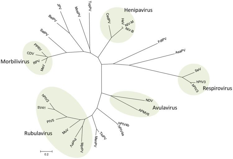
Diversity and evolution
In the past few decades, paramyxoviruses have been discovered from terrestrial, volant and aquatic animals, demonstrating a vast host range and great viral genetic diversity. As molecular technology advances and viral surveillance programs are implemented, the discovery of new viruses in this group is increasing.
The evolution of paramyxoviruses is still debated. Using pneumoviruses (mononegaviral family Pneumoviridae as an outgroup, paramyxoviruses can be divided into two clades: one consisting of avulaviruses and rubulaviruses and one consisting of respiroviruses, henipaviruses, and morbilliviruses. Within the second clade the respiroviruses appear to be the basal group. The respirovirus-henipavirus-morbillivirus may be basal to the avulavirus-rubulavirus clade.