
 | ||
The N-methyl-D-aspartate receptor (also known as the NMDA receptor or NMDAR), is a glutamate receptor and ion channel protein found in nerve cells. It is activated when glutamate and glycine (or D-serine) bind to it, and when activated it allows positively charged ions to flow through the cell membrane. The NMDA receptor is very important for controlling synaptic plasticity and memory function.
Contents
- Structure
- GluN1
- GluN2
- NR2B to NR2C switch
- The Role of NMDA Receptors in Excitotoxicity
- Differing Cascade Pathways
- Neural Plasticity
- The Role of Differing Subunits
- Excitotoxicity in a Clinical Setting
- Agonists
- Partial agonists
- Antagonists
- Modulators
- Receptor modulation
- Clinical significance
- References
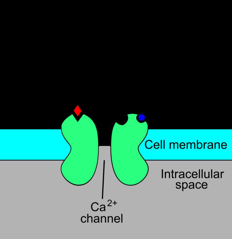
The NMDAR is a specific type of ionotropic glutamate receptor. The NMDA receptor is named this because the agonist molecule N-methyl-D-aspartate (NMDA) binds selectively to it, and not to other glutamate receptors. Activation of NMDA receptors results in the opening of an ion channel that is nonselective to cations, with a reversal potential near 0 mV. While the opening and closing of the ion channel is primarily gated by ligand binding, the current flow through the ion channel is voltage dependent. Extracellular magnesium (Mg2+) and zinc (Zn2+) ions can bind to specific sites on the receptor, blocking the passage of other cations through the open ion channel. Depolarization of the cell dislodges and repels the Mg2+ and Zn2+ ions from the pore, thus allowing a voltage-dependent flow of sodium (Na+) and small amounts of calcium (Ca2+) ions into the cell and potassium (K+) out of the cell.
Ca2+ flux through NMDARs is thought to be critical in synaptic plasticity, a cellular mechanism for learning and memory. The opening and closing (gating) of the NMDA receptor is complex. While it is primarily a ligand-gated channel, it does display weaker voltage-dependence modulation of the ligand-dependent gating. The ligand gating requires co-activation by two ligands: glutamate and either D-serine or glycine. The voltage-dependence of current through the channel is mainly due to binding of Mg2+ or Zn2+ ions to the protein as described above.
The activity of the NMDA receptor is affected by many psychoactive drugs such as phencyclidine (PCP), alcohol (ethanol) and dextromethorphan (DXM). The anaesthetic effects of the drugs ketamine and nitrous oxide are partially because of their effects on NMDA receptor activity.
Structure
The NMDA receptor forms a heterotetramer between two GluN1 and two GluN2 subunits (the subunits were previously denoted as NR1 and NR2), two obligatory NR1 subunits and two regionally localized NR2 subunits. A related gene family of NR3 A and B subunits have an inhibitory effect on receptor activity. Multiple receptor isoforms with distinct brain distributions and functional properties arise by selective splicing of the NR1 transcripts and differential expression of the NR2 subunits.
Each receptor subunit has modular design and each structural module also represents a functional unit:
The glycine-binding modules of the NR1 and NR3 subunits and the glutamate-binding module of the NR2A subunit have been expressed as soluble proteins, and their three-dimensional structure has been solved at atomic resolution by x-ray crystallography. This has revealed a common fold with amino acid-binding bacterial proteins and with the glutamate-binding module of AMPA-receptors and kainate-receptors.
GluN1
There are eight variants of the NR1 subunit produced by alternative splicing of GRIN1:
GluN2
While a single NR2 subunit is found in invertebrate organisms, four distinct isoforms of the NR2 subunit are expressed in vertebrates and are referred to with the nomenclature NR2A through NR2D (encoded by GRIN2A, GRIN2B, GRIN2C, GRIN2D). Strong evidence shows that the genes encoding the NR2 subunits in vertebrates have undergone at least two rounds of gene duplication. They contain the binding-site for the neurotransmitter glutamate. More importantly, each NR2 subunit has a different intracellular C-terminal domain that can interact with different sets of signalling molecules. Unlike NR1 subunits, NR2 subunits are expressed differentially across various cell types and control the electrophysiological properties of the NMDA receptor. One particular subunit, NR2B, is mainly present in immature neurons and in extrasynaptic locations, and contains the binding-site for the selective inhibitor ifenprodil.
Whereas NR2B is predominant in the early postnatal brain, the number of NR2A subunits grows, and eventually NR2A subunits outnumber NR2B. This is called the NR2B-NR2A developmental switch, and is notable because of the different kinetics each NR2 subunit lends to the receptor. For instance, greater ratios of the NR2B subunit leads to NMDA receptors which remain open longer compared to those with more NR2A. This may in part account for greater memory abilities in the immediate postnatal period compared to late in life, which is the principle behind genetically altered 'doogie mice'.
There are three hypothetical models to describe this switch mechanism:
The NR2B and NR2A subunits also have differential roles in mediating excitotoxic neuronal death. The developmental switch in subunit composition is thought to explain the developmental changes in NMDA neurotoxicity. Disruption of the gene for NR2B in mice causes perinatal lethality, whereas the disruption of NR2A gene produces viable mice, although with impaired hippocampal plasticity. One study suggests that reelin may play a role in the NMDA receptor maturation by increasing the NR2B subunit mobility.
NR2B to NR2C switch
Granule cell precursors (GCPs) of the cerebellum, after undergoing symmetric cell division in the external granule-cell layer (EGL), migrate into the internal granule-cell layer (IGL) where they downregulate NR2B and activate NR2C, a process that is independent of neuregulin beta signaling through ErbB2 and ErbB4 receptors.
The Role of NMDA Receptors in Excitotoxicity
NMDA receptors have been implicated by a number of studies to be strongly involved with excitotoxicity. Because NMDA receptors play an important role in the health and function of neurons, there has been much discussion on how these receptors can affect both cell survival and cell death. Recent evidence supports the hypothesis that overstimulation of extrasynaptic NMDA receptors has more to do with excitotoxicity than stimulation of their synaptic counterparts. In addition, while stimulation of extrasynaptic NMDA receptors appear to contribute to cell death, there is evidence to suggest that stimulation of synaptic NDMA receptors contributes to the health and longevity of the cell. There is ample evidence to support the dual nature of NMDA receptors based on location, and the hypothesis explaining the two differing mechanisms is known as the "Localization Hypothesis".
Differing Cascade Pathways
In order to support the localization hypothesis, it would be necessary to show differing cellular signaling pathways are activated by NMDA receptors based on its location within the cell membrane. Experiments have been designed to stimulate either synaptic or non-synaptic NMDA receptors exclusively. These types of experiments have shown that different pathways are being activated or regulated depending on the location of the signal origin. Many of these pathways use the same protein signals, but are regulated oppositely by NMDARs depending on its location. For example, synaptic NMDA excitation caused a decrease in the intracellular concentration of p38 mitogen-activated protein kinase (p38MAPK). Extrasynaptic stimulation NMDARs regulated p38MAPK in the opposite fashion, causing an increase in intracellular concentration. Experiments of this type have since been repeated with the results indicating these differences stretch across many pathways linked to cell survival and excitotoxicity.
Two specific proteins have been identified as a major pathway responsible for these different cellular responses ERK1/2, and Jacob. ERK1/2 is responsible for phosphorylation of Jacob when excited by synaptic NMDARs. This information is then transported to the nucleus. Phosphorylation of Jacob does not take place with extrasynaptic NMDA stimulation. This allows the transcription factors in the nucleus to respond differently based in the phosphorylation state of Jacob.
Neural Plasticity
NMDA receptors are also associated with synaptic plasticity. The idea that both synaptic and extrasynaptic NMDA receptors can affect long-term potentiation (LTP) and long-term depression (LTD) differently has also been explored. Experimental data suggest that extrasynaptic NMDA receptors inhibit LTP while producing LTD. Inhibition of LTP can be prevented with the introduction of a NMDA antagonist. A theta burst stimulation that usually induces LTP with synaptic NMDARs, when applied selectively to extrasynaptic NMDARs produces a LTD. Experimentation also indicates that extrasynaptic activity is not required for the formation of LTP. In addition, both synaptic and extrasynaptic are involved in expressing a full LTD.
The Role of Differing Subunits
Another factor that seems to affect NMDAR induced toxicity is the observed variation in subunit makeup. NMDA receptors are heterotetramers with two GluN1 subunits and two variable subunits. Two of these variable subunits, GluN2A and GluN2B, have been shown to preferentially lead to cell survival and cell death cascades respectively. Although both subunits are found in synaptic and extrasynaptic NMDARs there is some evidence to suggest that the GluN2B subunit occurs more frequently in extrasynaptic receptors. This observation could help explain the dualistic role that NMDA receptors play in excitotoxicity.
Despite the compelling evidence and the relative simplicity of these two theories working in tandem, there is still disagreement about the significance of these claims. Some problems in proving these theories arise with the difficulty of using pharmacological means to determine the subtypes of specific NMDARs. In addition, the theory of subunit variation does not explain how this effect might predominate, as it is widely held that the most common tetramer, made from two GluN1 subunits and one of each subunit GluN2A and GluN2B, makes up a high percentage of the NMDARs.
Excitotoxicity in a Clinical Setting
Excitotoxicity has been thought to play a role in the degenerative properties of neurodegenerative conditions since the late 1950s. NMDA receptors seem to play an important role in many of these degenerative diseases affecting the brain. Most notably exocytotic events involving NMDA receptors have been linked to Alzheimer's Disease and Huntington's Disease as well as with other medical conditions such as strokes and epilepsy. Treating these conditions with one of the many known NMDA receptor antagonists, however, lead to a variety of unwanted side effects, some of which can be quite severe. These side effects are, in part, observed because the NMDA receptors do not just signal for cell death but also play an important role in its vitality. Treatment for these conditions might be found in blocking NDMA receptors not found at the synapse.
Agonists
Activation of NMDA receptors requires binding of glutamate or aspartate (aspartate does not stimulate the receptors as strongly). In addition, NMDARs also require the binding of the co-agonist glycine for the efficient opening of the ion channel, which is a part of this receptor.
D-serine has also been found to co-agonize the NMDA receptor with even greater potency than glycine. D-Serine is produced by serine racemase, and is enriched in the same areas as NMDA receptors. Removal of D-serine can block NMDA-mediated excitatory neurotransmission in many areas. Recently, it has been shown that D-serine can be released both by neurons and astrocytes to regulate NMDA receptors.
NMDA receptor (NMDAR)-mediated currents are directly related to membrane depolarization. NMDA agonists therefore exhibit fast Mg2+ unbinding kinetics, increasing channel open probability with depolarization. This property is fundamental to the role of the NMDA receptor in memory and learning, and it has been suggested that this channel is a biochemical substrate of Hebbian learning, where it can act as a coincidence detector for membrane depolarization and synaptic transmission.
Some known NMDA receptor agonists include:
Partial agonists
Glycine-site NMDA receptor partial agonists, such as rapastinel and apimostinel, are now viewed for the development of new drugs with antidepressant and analgesic effects without obvious psychotomimetic activities.
Antagonists
Antagonists of the NMDA receptor are used as anesthetics for animals and sometimes humans, and are often used as recreational drugs due to their hallucinogenic properties, in addition to their unique effects at elevated dosages such as dissociation. When certain NMDA receptor antagonists are given to rodents in large doses, they can cause a form of brain damage called Olney's lesions. NMDA receptor antagonists that have been shown to induce Olney's lesions include ketamine, phencyclidine, and dextrorphan (a metabolite of dextromethorphan), as well as some NMDA receptor antagonists used only in research environments. So far, the published research on Olney's lesions is inconclusive in its occurrence upon human or monkey brain tissues with respect to an increase in the presence of NMDA receptor antagonists.
Common agents in which NMDA receptor antagonism is the primary mechanism of action:
Some common agents in which weak NMDA receptor antagonism is a secondary or additional action include:
Kynurenic acid is an endogenous NMDA receptor antagonist.
Modulators
The NMDA receptor is modulated by a number of endogenous and exogenous compounds:
Receptor modulation
The NMDA receptor is a non-specific cation channel that can allow the passage of Ca2+ and Na+ into the cell and K+ out of the cell. The excitatory postsynaptic potential (EPSP) produced by activation of an NMDA receptor increases the concentration of Ca2+ in the cell. The Ca2+ can in turn function as a second messenger in various signaling pathways. However, the NMDA receptor cation channel is blocked by Mg2+ at resting membrane potential. Magnesium unblock is not instantaneous, to unblock all available channels, the postsynaptic cell must be depolarized for a sufficiently long period of time (in the scale of milliseconds).
Therefore, the NMDA receptor functions as a "molecular coincidence detector". Its ion channel opens only when the following two conditions are met: glutamate is bound to the receptor, and the postsynaptic cell is depolarized (which removes the Mg2+ blocking the channel). This property of the NMDA receptor explains many aspects of long-term potentiation (LTP) and synaptic plasticity.
NMDA receptors are modulated by a number of endogenous and exogenous compounds and play a key role in a wide range of physiological (e.g., memory) and pathological processes (e.g., excitotoxicity).
Clinical significance
Memantine is approved by the U.S. F.D.A and the European Medicines Agency for treatment of moderate-to-severe Alzheimer's disease, and has now received a limited recommendation by the UK's National Institute for Health and Care Excellence for patients who fail other treatment options.
Cochlear NMDARs are the target of intense research to find pharmacological solutions to treat tinnitus. Recently, NMDARs were associated with a rare autoimmune disease, anti-NMDAR encephalitis, that usually occurs due to cross reactivity of antibodies produced by the immune system against ectopic brain tissues, such as those found in teratoma.
NMDAR ligands, including ketamine, esketamine, rapastinel (GLYX-13), apimostinel (NRX-1074), 4-chlorokynurenine (AV-101), and CERC-301, are under development for the treatment of mood disorders, including major depressive disorder and treatment-resistant depression. In addition, ketamine is already employed for this purpose as an off-label therapy in some clinics.
Compared to dopaminergic stimulants, phencyclidine can produce a wider range of symptoms that resemble schizophrenia in healthy volunteers, in what has led to the glutamate hypothesis of schizophrenia. Experiments in which rodents are treated with NMDA receptor antagonist are today the most common model when it comes to testing of novel schizophrenia therapies or exploring the exact mechanism of drugs already approved for treatment of schizophrenia.