
Entrez 4137 | Ensembl n/a | |
 | ||
Aliases MAPT, DDPAC, FTDP-17, MAPTL, MSTD, MTBT1, MTBT2, PPND, PPP1R103, TAU, microtubule associated protein tau, Tau proteins External IDs OMIM: 157140 MGI: 97180 HomoloGene: 74962 GeneCards: MAPT | ||
Tau proteins (or τ proteins, after the Greek letter with that name) are proteins that stabilize microtubules. They are abundant in neurons of the central nervous system and are less common elsewhere, but are also expressed at very low levels in CNS astrocytes and oligodendrocytes. Pathologies and dementias of the nervous system such as Alzheimer's disease and Parkinson's disease are associated with tau proteins that have become defective and no longer stabilize microtubules properly.
Contents
- Function
- Structure
- Genetics
- Clinical significance
- Traumatic brain injury
- Tau hypothesis of Alzheimers disease
- Interactions
- References
The tau proteins are the product of alternative splicing from a single gene that in humans is designated MAPT (microtubule-associated protein tau) and is located on chromosome 17.
The tau proteins were identified in 1975 as heat stable proteins essential for microtubule assembly and have been shown to have the characteristics of natively unfolded proteins.
Function
Tau protein is a highly soluble microtubule-associated protein (MAP). In humans, these proteins are found mostly in neurons compared to non-neuronal cells. One of tau's main functions is to modulate the stability of axonal microtubules. Other nervous system MAPs may perform similar functions, as suggested by tau knockout mice that did not show abnormalities in brain development - possibly because of compensation in tau deficiency by other MAPs. Tau is not present in dendrites and is active primarily in the distal portions of axons where it provides microtubule stabilization but also flexibility as needed. This contrasts with MAP6 (STOP) proteins in the proximal portions of axons, which, in essence, lock down the microtubules and MAP2 that stabilizes microtubules in dendrites.
Tau proteins interact with tubulin to stabilize microtubules and promote tubulin assembly into microtubules. Tau has two ways of controlling microtubule stability: isoforms and phosphorylation.
Structure
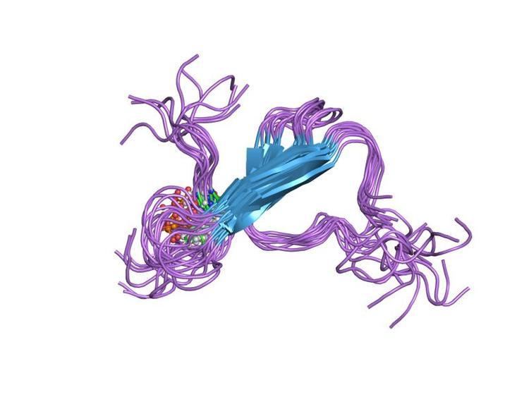
Six tau isoforms exist in human brain tissue, and they are distinguished by their number of binding domains. Three isoforms have three binding domains and the other three have four binding domains. The binding domains are located in the carboxy-terminus of the protein and are positively charged (allowing it to bind to the negatively charged microtubule). The isoforms with four binding domains are better at stabilizing microtubules than those with three binding domains. The isoforms are a result of alternative splicing in exons 2, 3, and 10 of the tau gene.
Tau is a phosphoprotein with 79 potential Serine (Ser) and Threonine (Thr) phosphorylation sites on the longest tau isoform. Phosphorylation has been reported on approximately 30 of these sites in normal tau proteins.
Phosphorylation of tau is regulated by a host of kinases, including PKN, a serine/threonine kinase. When PKN is activated, it phosphorylates tau, resulting in disruption of microtubule organization.
Phosphorylation of tau is also developmentally regulated. For example, fetal tau is more highly phosphorylated in the embryonic CNS than adult tau. The degree of phosphorylation in all six isoforms decreases with age due to the activation of phosphatases. Like kinases, phosphatases too play a role in regulating the phosphorylation of tau. For example, PP2A and PP2B are both present in human brain tissue and have the ability to dephosphorylate Ser396. The binding of these phosphatases to tau affects tau's association with MTs.
Genetics
In humans, the MAPT gene for encoding tau protein is located on chromosome 17q21, containing 16 exons. The major tau protein in the human brain is encoded by 11 exons. Exons 2, 3 and 10 are alternatively spliced, allowing six combinations (2–3–10–; 2+3–10–; 2+3+10–; 2–3–10+; 2+3–10+; 2+3+10+). Thus, in the human brain, the tau proteins constitute a family of six isoforms with the range from 352-441 amino acids. They differ in either zero, one, or two inserts of 29 amino acids at the N-terminal part (exon 2 and 3), and three or four repeat-regions at the C-terminal part (exon 10). So, the longest isoform in the CNS has four repeats (R1, R2, R3 and R4) and two inserts (441 amino acids total), while the shortest isoform has three repeats (R1, R3 and R4) and no insert (352 amino acids total).
The MAPT gene has two haplogroups, H1 and H2, in which the gene appears in inverted orientations. Haplogroup H2 is common only in Europe and in people with European ancestry. Haplogroup H1 appears to be associated with increased probability of certain dementias, such as Alzheimer's disease. The presence of both haplogroups in Europe means that recombination between inverted haplotypes can result in the lack of one of the functioning copy of the gene, resulting in congenital defects.
Clinical significance
Hyperphosphorylation of the tau protein (tau inclusions, pTau) can result in the self-assembly of tangles of paired helical filaments and straight filaments, which are involved in the pathogenesis of Alzheimer's disease, frontotemporal dementia, and other tauopathies.
All of the six tau isoforms are present in an often hyperphosphorylated state in paired helical filaments from Alzheimer's disease brain. In other neurodegenerative diseases, the deposition of aggregates enriched in certain tau isoforms has been reported. When misfolded, this otherwise very soluble protein can form extremely insoluble aggregates that contribute to a number of neurodegenerative diseases.
Recent research suggests that tau may be released extracellularly by an exosome-based mechanism in Alzheimer's disease.
Gender-specific tau gene expression across different regions of the human brain has recently been implicated in gender differences in the manifestations and risk for tauopathies.
Some aspects of how the disease functions also suggest that it has some similarities to prion proteins.
Traumatic brain injury
Repetitive mild traumatic brain injury (TBI), now recognized as a central component of brain injury in contact sports, especially American football, and the concussive force of military blasts. can lead to chronic traumatic encephalopathy (CTE) that is characterized by fibrillar tangles of hyperphosphorylated tau.
High levels of tau protein in fluid bathing the brain are linked to poor recovery after head trauma.
Tau hypothesis of Alzheimer's disease
The tau hypothesis states that excessive or abnormal phosphorylation of tau results in the transformation of normal adult tau into PHF-tau (paired helical filament) and NFTs (neurofibrillary tangles). Tau protein is a highly soluble microtubule-associated protein (MAP). Through its isoforms and phosphorylation tau protein interacts with tubulin to stabilize microtubule assembly. Tau proteins constitute a family of six isoforms with the range from 352-441 amino acids. The longest isoform in the CNS has four repeats (R1, R2, R3, and R4) and two inserts (441 amino acids total), whereas the shortest isoform has three repeats (R1, R3, and R4) and no insert (352 amino acids total). All of the six tau isoforms are present in an often hyperphosphorylated state in paired helical filaments from AD.
Mutations that alter function and isoform expression of tau lead to hyperphosphorylation. The process of tau aggregation in the absence of mutations is not known but might result from increased phosphorylation, protease action or exposure to polyanions, such as glycosaminoglycans.[6] Hyperphosphorylated tau disassembles microtubules and sequesters normal tau, MAP 1(microtubule associated protein1), MAP 2, and ubiquitin into tangles of PHFs. This insoluble structure damages cytoplasmic functions and interferes with axonal transport, which can lead to cell death.
Interactions
Tau protein has been shown to interact with proto-oncogene tyrosine-protein kinase: