
 | ||
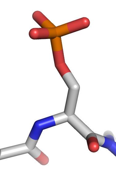
Phosphorylation is the addition of a phosphoryl group (PO3)− to a molecule. In biology, phosphorylation and its counterpart, dephosphorylation, are critical for many cellular processes. A large fraction of proteins are at least temporarily phosphorylated, as are many sugars, lipids, and other molecules. Phosphorylation is especially important for protein function as this modification activates (or deactivates) many enzymes, thereby regulating their function. Protein phosphorylation is one type of post-translational modification.
Contents
- Phosphorylation of glucose
- Function
- Signaling networks
- Types of phosphorylation
- Phosphorylation of histones
- Detection and characterization
- ATP
- References
The prominent role of protein phosphorylation in biochemistry is illustrated by the huge body of studies published on the subject (as of March 2015, the MEDLINE database returns over 240,000 articles, mostly on protein phosphorylation).
Phosphorylation of glucose
Phosphorylation of sugars is often the first stage of their catabolism. It allows cells to accumulate sugars because the phosphate group prevents the molecules from diffusing back across their transporter. Phosphorylation of glucose is a key reaction in sugar metabolism because many sugars are first converted to glucose before they are metabolized further.
The chemical equation for the conversion of D-glucose to D-glucose-6-phosphate in the first step of glycolysis is given by D-glucose + ATP -> D-glucose-6-phosphate + ADP ΔG° = -16.7 kJ/mol.
Research D. G. Walker of the University of Birmingham determined the presence of two specific enzymes in adult guinea pig liver, both of which catalyze the phosphorylation of glucose to glucose 6 phosphate. The two enzymes have been identified as a specific glucokinase (ATP-D-glucose 6-phosphotransferase) and non-specific hexokinase (ATP-D-hexose 6-phosphotransferase).
Hepatic cell is freely permeable to glucose, and the initial rate of phosphorylation of glucose is the rate-limiting step in glucose metabolism by the liver (ATP-D-glucose 6-phosphotransferase) and non-specific hexokinase (ATP-D-hexose 6-phosphotransferase).
The role of glucose 6-phosphate in glycogen synthase: High blood glucose concentration causes an increase in intracellular levels of glucose 6 phosphate in liver, skeletal muscle and fat (adipose) tissue. (ATP-D-glucose 6-phosphotransferase) and non-specific hexokinase (ATP-D-hexose 6-phosphotransferase). In liver, synthesis of glycogen is directly correlated by blood glucose concentration and in skeletal muscle and adipocytes, glucose has a minor effect on glycogen synthase. High blood glucose releases insulin, stimulating the trans location of specific glucose transporters to the cell membrane.
The liver’s crucial role in controlling blood sugar concentrations by breaking down glucose into carbon dioxide and glycogen is characterized by the negative delta G value, which indicates that this is a point of regulation with. The hexokinase enzyme has a low Km, indicating a high affinity for glucose, so this initial phosphorylation can proceed even when glucose levels at nanoscopic scale within the blood.
The phosphorylation of glucose can be enhanced by the binding of Fructose-6-phosphate, and lessened by the binding fructose-1-phosphate. Fructose consumed in the diet is converted to F1P in the liver. This negates the action of F6P on glucokinase, which ultimately favors the forward reaction. The capacity of liver cells to phosphorylate fructose exceeds capacity of metabolize fructose-1-phosphate. Consuming excess fructose ultimately results in an imbalance in liver metabolism, which indirectly exhausts the liver cell’s supply of ATP.
Allosteric activation by glucose 6 phosphate, which acts as an effector, stimulates glycogen synthase, and glucose 6 phosphate may inhibit the phosphorylation of glycogen synthase by cyclic AMP-stimulated protein kinase.
Phosphorylation of glucose is imperative in processes within the body. For example, phosphorylating glucose is necessary for insulin-dependent mechanistic target of rapamycin pathway activity within the heart. This further suggests a link between intermediary metabolism and cardiac growth.
Function
Reversible phosphorylation of proteins is an important regulatory mechanism that occurs in both prokaryotic and eukaryotic organisms. Kinases phosphorylate proteins and phosphatases dephosphorylate proteins. Many enzymes and receptors are switched "on" or "off" by phosphorylation and dephosphorylation. Reversible phosphorylation results in a conformational change in the structure in many enzymes and receptors, causing them to become activated or deactivated. Phosphorylation usually occurs on serine, threonine, tyrosine and histidine residues in eukaryotic proteins. Histidine phosphorylation of eukaryotic proteins appears to be much more frequent than tyrosine phosphorylation. In prokaryotic proteins phosphorylation occurs on the serine, threonine, tyrosine, histidine or arginine or lysine residues. The addition of a phosphate (PO4) molecule to a polar R group of an amino acid residue can turn a hydrophobic portion of a protein into a polar and extremely hydrophilic portion of a molecule. In this way protein dynamics can induce a conformational change in the structure of the protein via long-range allostery with other hydrophobic and hydrophilic residues in the protein.
One such example of the regulatory role that phosphorylation plays is the p53 tumor suppressor protein. The p53 protein is heavily regulated and contains more than 18 different phosphorylation sites. Activation of p53 can lead to cell cycle arrest, which can be reversed under some circumstances, or apoptotic cell death. This activity occurs only in situations wherein the cell is damaged or physiology is disturbed in normal healthy individuals.
Upon the deactivating signal, the protein becomes dephosphorylated again and stops working. This is the mechanism in many forms of signal transduction, for example the way in which incoming light is processed in the light-sensitive cells of the retina.
Regulatory roles of phosphorylation include
Glycolysis is an essential process of glucose degrading into two molecules of pyruvate, through various steps, with the help of different enzymes. It occurs in ten steps and proves that phosphorylation is a much required and necessary to attain the end products. Phosphorylation initiates the reaction in step 1 of the preparatory step (first half of glycolysis), and initiates step 6 of payoff phase (second phase of glycolysis).
Glucose, by nature, is a small molecule with the ability to diffuse in and out of the cell. By phosphorylating glucose (adding a negatively charged phosphate group), glucose is converted to glucose-6-phosphate and trapped within the cell as the cell membrane is negatively charged. This reaction occurs due to the enzyme hexokinase, an enzyme that helps phosphorylate many six-membered ring structures. Glucose-6-phosphate cannot travel through the cell membrane and is therefore, coerced to stay inside the cell. Phosphorylation takes place in step 3, where fructose-6-phosphate is converted to fructose-1,6-bisphosphate. This reaction is catalyzed by phosphofructokinase.
While phosphorylation is performed by ATPs during preparatory steps, phosphorylation during payoff phase is maintained by inorganic phosphate. Each molecule of glyceraldehyde-3-phosphate is phosphorylated to form 1,3-bisphosphoglycerate. This reaction is catalyzed by GAPDH (glyceraldehyde-3-phosphate dehydrogenase). The cascade effect of phosphorylation eventually causes instability and allows enzymes to open the carbon bonds in glucose.
Phosphorylation functions as an extremely vital component of glycolysis, for it helps in transport, control and efficiency.
Signaling networks
Elucidating complex signaling pathway phosphorylation events can be difficult. In cellular signaling pathways, protein A phosphorylates protein B, and B phosphorylates C. However, in another signaling pathway, protein D phosphorylates A, or phosphorylates protein C. Global approaches such as phosphoproteomics, the study of phosphorylated proteins, which is a sub-branch of proteomics, combined with mass spectrometry-based proteomics, have been utilised to identify and quantify dynamic changes in phosphorylated proteins over time. These techniques are becoming increasingly important for the systematic analysis of complex phosphorylation networks. They have been successfully used to identify dynamic changes in the phosphorylation status of more than 6000 sites after stimulation with epidermal growth factor. Another approach for understanding Phosphorylation Network, is by measuring the genetic interactions between multiple phosphorylating proteins and their targets. This reveals interesting recurring patterns of interactions – network motifs. Computational methods have been developed to model phosphorylation networks and predict their responses under different perturbations.
Types of phosphorylation
Within a protein, phosphorylation can occur on several amino acids. Phosphorylation on serine is the most common, followed by threonine. Tyrosine phosphorylation is relatively rare but is at the origin of protein phosphorylation signaling pathways in most of the eukaryotes. Phosphorylation on amino acids, such as serine, threonine, and tyrosine results in the formation of a phosphoprotein, when the phosphate group of the phosphoprotein reacts with the -OH group of a Ser, Thr, or Tyr sidechain in an esterification reaction. However, since tyrosine phosphorylated proteins are relatively easy to purify using antibodies, tyrosine phosphorylation sites are relatively well understood. Histidine and aspartate phosphorylation occurs in prokaryotes as part of two-component signaling and in some cases in eukaryotes in some signal transduction pathways. In prokaryotes, archea, and some lower eukaryotes histidine's nitrogen act as a nucleophile and binds to a phosphate group. Once histidine is phosphorylated the regulatory domain of the response regulator catalyzes the transfer of the phosphate to aspartate.
Phosphorylation of histones
Eukaryotic DNA is organized with histone proteins in specific complexes called chromatin. The chromatin structure functions and facilitates the packaging, organization and distribution of eukaryotic DNA. However, it has a negative impact on several fundamental biological processes such as transcription, replication and DNA repair by restricting the accessibility of certain enzymes and proteins. Post-translational modification of histones such as histone phosphorylation has been shown to modify the chromatin structure by changing protein:DNA or protein:protein interactions. Histone post-translational modifications modify the chromatin structure. The most commonly associated histone phosphorylation occurs during cellular responses to DNA damage, when phosphorylated histone H2A separates large chromatin domains around the site of DNA breakage. Researchers investigated whether modifications of histones directly impact RNA polymerase II directed transcription. Researchers choose proteins that are known to modify histones to test their effects on transcription, and found that the stress-induced kinase, MSK1, inhibits RNA synthesis. Inhibition of transcription by MSK1 was most sensitive when the template was in chromatin, since DNA templates not in chromatin were resistant to the effects of MSK1. It was shown that MSK1 phosphorylated histone H2A on serine 1, and mutation of serine 1 to alanine blocked the inhibition of transcription by MSK1. Thus results suggested that the acetylation of histones can stimulate transcription by suppressing an inhibitory phosphorylation by a kinase as MSK1.
Detection and characterization
Antibodies can be used as powerful tool to detect whether a protein is phosphorylated at a particular site. Antibodies bind to and detect phosphorylation-induced conformational changes in the protein. Such antibodies are called phospho-specific antibodies; hundreds of such antibodies are now available. They are becoming critical reagents both for basic research and for clinical diagnosis.
Posttranslational modification (PTM) isoforms are easily detected on 2D gels. Indeed, phosphorylation replaces neutral hydroxyl groups on serines, threonines, or tyrosines with negatively charged phosphates with pKs near 1.2 and 6.5. Thus, below pH 5.5, phosphates add a single negative charge; near pH 6.5, they add 1.5 negative charges; above pH 7.5, they add 2 negative charges. The relative amount of each isoform can also easily and rapidly be determined from staining intensity on 2D gels.
In some very specific cases, the detection of the phosphorylation as a shift in the protein's electrophoretic mobility is possible on simple 1-dimensional SDS-PAGE gels, as it's described for instance for a transcriptional coactivator by Kovacs et al. Strong phosphorylation-related conformational changes (that persist in detergent-containing solutions) are thought to underlie this phenomenon. Most of the phosphorylation sites for which such a mobility shift has been described fall in the category of SP and TP sites (i.e. a proline residue follows the phosphorylated serine or threonine residue).
More recently large-scale mass spectrometry analyses have been used to determine sites of protein phosphorylation. Over the last 4 years, dozens of studies have been published, each identifying thousands of sites, many of which were previously undescribed. Mass spectrometry is ideally suited for such analyses using HCD or ETD fragmentation, as the addition of phosphorylation results in an increase in the mass of the protein and the phosphorylated residue. Advanced, highly accurate mass spectrometers are needed for these studies, limiting the technology to labs with high-end mass spectrometers. However, the analysis of phosphorylated peptides by mass spectrometry is still not as straightforward as for “regular”, unmodified peptides. Recently EThcD has been developed combining electron-transfer and higher-energy collision dissociation. Compared to the usual fragmentation methods, EThcD scheme provides more informative MS/MS spectra for unambiguous phosphosite localization.
A detailed characterization of the sites of phosphorylation is very difficult, and the quantitation of protein phosphorylation by mass spectrometry requires isotopic internal standard approaches. A relative quantitation can be obtained with a variety of differential isotope labeling technologies. There are also several quantitative protein phosphorylation methods, including fluorescence immunoassays, Microscale thermophoresis, FRET, TRF, fluorescence polarization, fluorescence-quenching, mobility shift, bead-based detection, and cell-based formats.
ATP
ATP, the "high-energy" exchange medium in the cell, is synthesized in the mitochondrion by addition of a third phosphate group to ADP in a process referred to as oxidative phosphorylation. ATP is also synthesized by substrate-level phosphorylation during glycolysis. ATP is synthesized at the expense of solar energy by photophosphorylation in the chloroplasts of plant cells.