
Specialty pulmonology ICD-9-CM 495 MedlinePlus 000109 | ICD-10 J67 DiseasesDB 4630 eMedicine med/1103 ped/2577 | |
 | ||
Hypersensitivity pneumonitis (HP; also called allergic alveolitis or extrinsic allergic alveolitis, EAA) is an inflammation of the alveoli within the lung caused by hypersensitivity to inhaled organic dusts. Sufferers are commonly exposed to the dust by their occupation or hobbies.
Contents
Signs and symptoms
Hypersensitivity pneumonitis (HP) is categorized as acute, subacute, and chronic based on the duration of the illness.
Acute
In the acute form of HP, symptoms may develop 4–6 hours following heavy exposure to the provoking antigen. Symptoms include fever, chills, malaise, cough, chest tightness, dyspnea, rash, swelling and headache. Symptoms resolve within 12 hours to several days upon cessation of exposure.
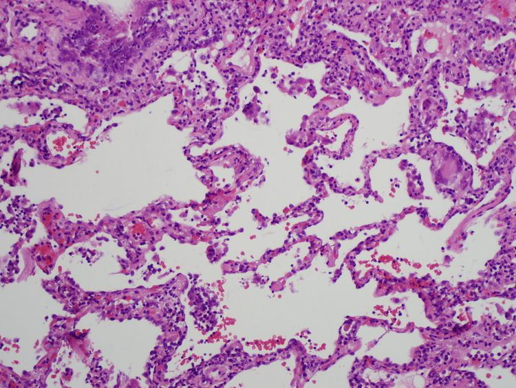
Acute HP is characterized by poorly formed noncaseating interstitial granulomas and mononuclear cell infiltration in a peribronchial distribution with prominent giant cells.
On chest radiographs, a diffuse micronodular interstitial pattern (at times with ground-glass density in the lower and middle lung zones) may be observed. Findings are normal in approximately 10% of patients." In high-resolution CT scans, ground-glass opacities or diffusely increased radiodensities are present. Pulmonary function tests show reduced diffusion capacity of lungs for carbon monoxide (DLCO). Many patients have hypoxemia at rest, and all patients desaturate with exercise. Extrinsic allergic alveolitis may eventually lead to Interstitial lung disease.
Subacute
Patients with subacute HP gradually develop a productive cough, dyspnea, fatigue, anorexia, weight loss, and pleurisy. Symptoms are similar to the acute form of the disease, but are less severe and last longer. On chest radiographs, micronodular or reticular opacities are most prominent in mid-to-lower lung zones. Findings may be present in patients who have experienced repeated acute attacks.
The subacute, or intermittent, form produces more well-formed noncaseating granulomas, bronchiolitis with or without organizing pneumonia, and interstitial fibrosis.
Chronic
In chronic HP, patients often lack a history of acute episodes. They have an insidious onset of cough, progressive dyspnea, fatigue, and weight loss. This is associated with partial to complete but gradual reversibility. Avoiding any further exposure is recommended. Clubbing is observed in 50% of patients. Tachypnea, respiratory distress, and inspiratory crackles over lower lung fields often are present.
On chest radiographs, progressive fibrotic changes with loss of lung volume particularly affect the upper lobes. Nodular or ground-glass opacities are not present. Features of emphysema are found on significant chest films and CT scans.
Chronic forms reveal additional findings of chronic interstitial inflammation and alveolar destruction (honeycombing) associated with dense fibrosis. Cholesterol clefts or asteroid bodies are present within or outside granulomas.
In addition, many patients have hypoxemia at rest, and all patients desaturate with exercise.
Pathophysiology
Hypersensitivity pneumonitis involves inhalation of an antigen. This leads to an exaggerated immune response (hypersensitivity). Type III hypersensitivity and type IV hypersensitivity can both occur depending on the cause.
Diagnosis
The diagnosis is based upon a history of symptoms after exposure to the allergen and clinical tests. A physician may take blood tests, seeking signs of inflammation, a chest X-ray and lung function tests. The sufferer shows a restrictive loss of lung function.
Precipitating IgG antibodies against fungal or avian antigens can be detected in the laboratory using the traditional Ouchterlony immunodiffusion method wherein 'precipitin' lines form on agar plate. The ImmunoCAP technology has replaced this time consuming, labor-intensive method with their automated CAP assays and FEIA (Fluorescence enzyme immunoassay) that can detect IgG antibodies against Aspergillus fumigatus (Farmer's lung or for ABPA) or avian antigens (Bird Fancier's Lung).
Although overlapping in many cases, hypersensitivity pneumonitis may be distinguished from occupational asthma in that it is not restricted to only occupational exposure, and that asthma generally is classified as a type I hypersensitivity. Unlike asthma, hypersensitivity pneumonitis targets lung alveoli rather than bronchi.
Lung biopsy
Lung biopsies can be diagnostic in cases of chronic hypersensitivity pneumonitis, or may help to suggest the diagnosis and trigger or intensify the search for an allergen. The main feature of chronic hypersensitivity pneumonitis on lung biopsies is expansion of the interstitium by lymphocytes accompanied by an occasional multinucleated giant cell or loose granuloma.
When fibrosis develops in chronic hypersensitivity pneumonitis, the differential diagnosis in lung biopsies includes the idiopathic interstitial pneumonias. This group of diseases includes usual interstitial pneumonia, non-specific interstitial pneumonia and cryptogenic organizing pneumonia, among others.
The prognosis of some idiopathic interstitial pneumonias, e.g. idiopathic usual interstitial pneumonia (i.e. idiopathic pulmonary fibrosis), are very poor and the treatments of little help. This contrasts the prognosis (and treatment) for hypersensitivity pneumonitis, which is generally fairly good if the allergen is identified and exposures to it significantly reduced or eliminated. Thus, a lung biopsy, in some cases, may make a decisive difference.
Types
Hypersensitivity pneumonitis may also be called many different names, based on the provoking antigen. These include:
Of these types, Farmer's Lung and Bird-Breeder's Lung are the most common. "Studies document 8-540 cases per 100,000 persons per year for farmers and 6000-21,000 cases per 100,000 persons per year for pigeon breeders. High attack rates are documented in sporadic outbreaks. Prevalence varies by region, climate, and farming practices. HP affects 0.4-7% of the farming population. Reported prevalence among bird fanciers is estimated to be 20-20,000 cases per 100,000 persons at risk."
Treatment
The best treatment is to avoid the provoking allergen, as chronic exposure can cause permanent damage. Corticosteroids such as prednisolone may help to control symptoms but may produce side-effects.