
ICD-10 J84.1 DiseasesDB 4815 | ICD-9-CM 515 | |
 | ||
Usual interstitial pneumonia (UIP) is a form of lung disease characterized by progressive scarring of both lungs. The scarring (fibrosis) involves the supporting framework (interstitium) of the lung. UIP is thus classified as a form of interstitial lung disease. The term "usual" refers to the fact that UIP is the most common form of interstitial fibrosis. "Pneumonia" indicates "lung abnormality", which includes fibrosis and inflammation. A term previously used for UIP in the British literature is cryptogenic fibrosing alveolitis, a term that has fallen out of favor since the basic underlying pathology is now thought to be fibrosis, not inflammation.
Contents
Signs and symptoms
The typical symptoms of UIP are progressive shortness of breath and cough for a period of months. In some patients, UIP is diagnosed only when a more acute disease supervenes and brings the patient to medical attention.
Causes
The cause of the scarring in UIP may be known (less commonly) or unknown (more commonly). Since the medical term for conditions of unknown cause is "idiopathic", the clinical term for UIP of unknown cause is idiopathic pulmonary fibrosis (IPF). Examples of known causes of UIP include systemic sclerosis/scleroderma, rheumatoid arthritis, asbestosis, and prolonged use of medications such as nitrofurantoin or amiodarone.
Diagnosis
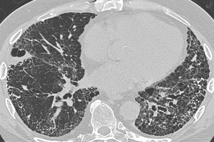
UIP may be diagnosed by a radiologist using computed tomography (CT) scan of the chest, or by a pathologist using tissue obtained by a lung biopsy. Radiologically, the main feature required for a confident diagnosis of UIP is honeycomb change in the periphery and the lower portions (bases) of the lungs. The histologic hallmarks of UIP, as seen in lung tissue under a microscope by a pathologist, are interstitial fibrosis in a "patchwork pattern", honeycomb change and fibroblast foci (see images below).
Differential diagnosis
The differential diagnosis includes other types of lung disease that cause similar symptoms and show similar abnormalities on chest radiographs. Some of these diseases cause fibrosis, scarring or honeycomb change. The most common considerations include:
Prognosis
Regardless of cause, UIP is relentlessly progressive, usually leading to respiratory failure and death. Some patients do well for a prolonged period of time, but then deteriorate rapidly because of a superimposed acute illness (so-called "accelerated UIP"). The outlook for long-term survival is poor. In most studies, the median survival is 3 to 4 years. Patients with UIP in the setting of rheumatoid arthritis have a slightly better prognosis than UIP without a known cause (IPF).
History
UIP, as a term, first appeared in the pathology literature. It was coined by Averill Abraham Liebow.