
Specialty medical genetics ICD-9-CM 753.5 DiseasesDB 33377 | ICD-10 Q64.1 OMIM 600057 eMedicine ped/704 | |
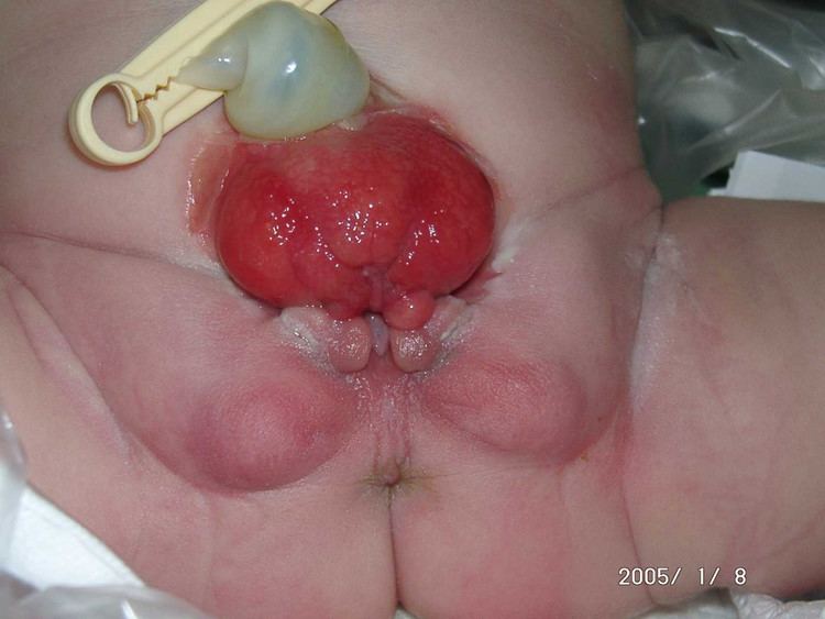 | ||
Bladder exstrophy (also known as Ectopia vesicae) is a congenital anomaly that exists along the spectrum of the exstrophy-epispadias complex and most notably involves protrusion of the urinary bladder through a defect in the abdominal wall. Its presentation is variable, often including abnormalities of the bony pelvis, pelvic floor, and genitalia. The underlying embryologic mechanism leading to bladder exstrophy is unknown, though it is thought to be in part due to failed reinforcement of the cloacal membrane by underlying mesoderm.
Contents
Incidence
Occurring at a rate between 1 in 10,000 to 1 in 50,000 with a male-to-female ratio of 2.3-6:1, bladder exstrophy is relatively rare. For those individuals with bladder exstrophy who maintain their ability to reproduce, the risk of bladder exstrophy in their children is approximately 500-fold greater than the general population.
Presentation
The classic manifestation of bladder exstrophy presents with:
Females frequently have a displaced and narrowed vaginal orifice, a bifid clitoris, and divergent labia.
Prenatal diagnosis
In a small retrospective study of 25 pregnancies five factors were found to be strongly associated with a prenatal diagnosis of bladder exstrophy:
While a diagnosis of bladder exstrophy was made retrospectively in a majority of pregnancies, in only three cases was a prenatal diagnosis made.
Management at birth
Upon delivery, the exposed bladder is irrigated and a non-adherent film is placed to prevent as much contact with the external environment as possible. In the event the child was not born at a medical center with an appropriate exstrophy support team then transfer will likely follow. Upon transfer, or for those infants born at a medical center able to care for bladder exstrophy, imaging may take place in the first few hours of life prior to the child undergoing surgery.
Primary (immediate) closure is indicated only in those patients with a bladder of appropriate size, elasticity, and contractility as those patients are most likely to develop a bladder of adequate capacity after early surgical intervention.
Conditions that are absolute contraindications despite bladder adequacy include duplication of the penis or scrotum and significant bilateral hydronephrosis.
Treatment
Modern therapy is aimed at surgical reconstruction of the bladder and genitalia. Both males and females are born with this anomaly. Treatment is similar.
In males treatments have been: In the Modern Staged Repair of Exstrophy (MSRE) the initial step is closure of the abdominal wall, often requiring a pelvic osteotomy. This leaves the patient with penile epispadias and urinary incontinence. At approximately 2–3 years of age the patient then undergoes repair of the epispadias after testosterone stimulation. Finally, bladder neck repair usually occurs around the age of 4–5 years, though this is dependent upon a bladder with adequate capacity and, most importantly, an indication that the child is interested in becoming continent. In the Complete Primary Repair of Exstrophy (CPRE) the bladder closure is combined with an epispadias repair, in an effort to decrease costs and morbidity. This technique has, however, led to significant loss of penile and corporal tissue, particularly in younger patients.
In females treatment has included: Surgical reconstruction of the clitoris which is separated into two distinct bodies. Surgical reconstruction to correct the split of the mons, redefine the structure of the bladder neck and urethra. Vaginoplasty will correct the anteriorly displaced vagina. If the anus is involved, it is also repaired. Fertility remains and women who were born with bladder extrophy usually develop prolapse due to the weaker muscles of the pelvic floor.
Prognosis
The most important criterion for improving long-term prognosis is success of the initial closure. If a patient requires more than one closure their chance of continence drops off precipitously with each additional closure - at just two closures the chance of voiding continence is just 17%.
Complications
Even with successful surgery, patients may have long-term complications. Some of the most common include: