
 | ||
Nerve Pudendal nervePelvic splanchnic nervesInferior hypogastric plexus Latin urethra vagina; feminina (female); urethra masculina (male) Similar Urethrorrhagia, Urethral gland, Urethrotomy | ||
In anatomy, the urethra (from Greek οὐρήθρα – ourḗthrā) is a tube that connects the urinary bladder to the urinary meatus for the removal of fluids from the body. In male placental mammals, the urethra travels through the penis, and carries semen as well as urine. In female placental mammals, the urethra is shorter and emerges at the female external urethral orifice above the vaginal opening.
Contents
- Female
- Male
- Histology
- Physiology
- Sexual physiology
- Clinical significance
- Investigations
- Catheterization
- References
Female placental mammals use their urethra only for urinating, but male placental mammals use their urethra for both urination and ejaculation. The external urethral sphincter is a striated muscle that allows voluntary control over urination.
Female
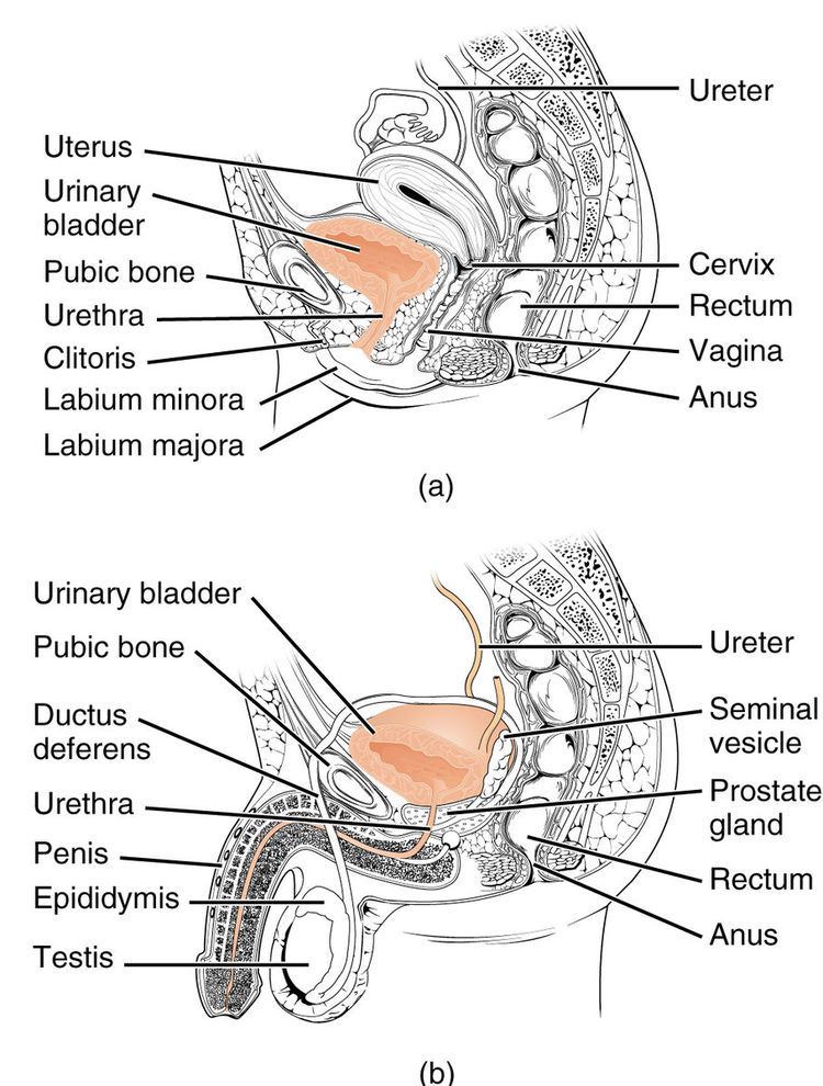
In the human female, the urethra is about 1.9 inches (4.8 cm) to 2 inches (5.1 cm) long and exits the body between the clitoris and the vagina, extending from the internal to the external urethral orifice. The meatus is located below the clitoris. It is placed behind the symphysis pubis, embedded in the anterior wall of the vagina, and its direction is obliquely downward and forward; it is slightly curved with the concavity directed forward. The proximal 2/3rds is lined by Transitional epithelium cells while distal 1/3rd is lined by stratified squamous Epithelium cells.
The urethra consists of three coats: muscular, erectile, and mucous, the muscular layer being a continuation of that of the bladder. Between the superior and inferior fascia of the urogenital diaphragm, the female urethra is surrounded by the urethral sphincter. Somatic (conscious) innervation of the external urethral sphincter is supplied by the pudendal nerve. The uro-genital sinus may be divided into three component parts. The first of these is the cranial portion which is continuous with the allantois and forms the bladder proper. The pelvic part of the sinus forms the prostatic urethra and epithelium as well as the membranous urethra and bulbo urethral glands in the male and the membranous urethra and part of the vagina in females.
Male
In the human male, the urethra is about 8 inches (20 cm) long and opens at the end of the external urethral meatus.{{}} The urethra provides an exit for urine as well as semen during ejaculation.
The urethra is divided into four parts in men, named after the location:
There is inadequate data for the typical length of the male urethra; however, a study of 109 men showed an average length of 22.3 cm (SD = 2.4 cm), ranging from 15 cm to 29 cm.
Histology
The epithelium of the urethra starts off as transitional cells as it exits the bladder. Further along the urethra there are pseudostratified columnar and stratified columnar epithelia, then stratified squamous cells near the external urethral orifice.
There are small mucus-secreting urethral glands, that help protect the epithelium from the corrosive urine.
Physiology
The urethra is the vessel through which urine passes after leaving the bladder. During urination, the smooth muscle lining the urethra relaxes in concert with bladder contraction(s) to forcefully expel the urine in a pressurized stream. Following this, the urethra re-establishes muscle tone by contracting the smooth muscle layer, and the bladder returns to a relaxed, quiescent state. Urethral smooth muscle cells are mechanically coupled to each other to coordinate mechanical force and electrical signaling in an organized, unitary fashion.
Sexual physiology
The male urethra is the conduit for semen during sexual intercourse. It also serves as a passage for urine to flow. Urine typically contains epithelial cells shed from the urinary tract. Urine cytology evaluates this urinary sediment for the presence of cancerous cells from the lining of the urinary tract, and it is a convenient noninvasive technique for follow-up analysis of patients treated for urinary tract cancers. For this process, urine must be collected in a reliable fashion, and if urine samples are inadequate, the urinary tract can be assessed via instrumentation. In urine cytology, collected urine is examined microscopically. One limitation is the inability to definitively identify low-grade cancer cells and urine cytology is used mostly to identify high-grade tumors.
Clinical significance
Investigations
As the urethra is an open vessel with a lumen, investigations of the genitourinary tract may involve the urethra. Endoscopy of the bladder may be conducted by the urethra, called cystoscopy.
Catheterization
During a hospital stay or surgical procedure, a catheter may be inserted through the urethra to drain urine from the bladder. The length of a male's urethra, and the fact it contains a prominent bend, makes catheterization more difficult. The integrity of the urethra can be determined by a procedure known as retrograde urethrogram.