
Precursor Ureteric bud MeSH A05.810.776 TA A08.2.01.001 | Latin Ureter Dorlands/Elsevier Ureter | |
 | ||
Artery Superior vesical artery, Vaginal artery, Ureteral branches of renal artery | ||
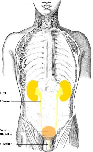
In human anatomy, the ureters are tubes made of smooth muscle fibers that propel urine from the kidneys to the urinary bladder. In the adult, the ureters are usually 25–30 cm (10–12 in) long and ~3–4 mm in diameter. Histologically, the ureter contains transitional epithelium and an additional smooth muscle layer in the more distal one-third to assist with peristalsis.
Contents
Structure
In humans, the ureters arise from the pelvis of each kidney, and descend on top of the psoas major muscle to reach the brim of the pelvis. Here, they cross in front of the common iliac arteries. They then pass down along the sides of the pelvis, and finally curve forwards and enter the bladder from its left and right sides at the back of the bladder. This is classically depicted as running "posteroinferiorly on the lateral walls of the pelvis and then curve anteromedially to enter the bladder". The orifices of the ureters are placed at the postero-lateral angles of the trigone of the bladder, and are usually slit-like in form. In the contracted bladder they are about 2.5 cm. apart and about the same distance from the internal urethral orifice; in the distended bladder these measurements may be increased to about 5 cm.
The junction between the pelvis of the kidney and the ureters is known as the ureteropelvic junction or ureteral pelvic junction, and the junction between the ureter and the bladder is known as the ureterovesical (ureter-bladder) junction. At the entrance to the bladder, the ureters are surrounded by valves known as ureterovesical valves, which prevent vesicoureteral reflux (backflow of urine).
In females, the ureters pass through the mesometrium and under the uterine arteries on the way to the urinary bladder.
Constrictions
The ureter has a diameter of 3 mm but there are five constrictions,]:
Development
During the embryologic development of the kidney, the ureteropelvic junction is the last part of the ureter to become patent.
Blood supply
The ureters receive a segmental arterial supply, which varies along its course.
- The upper part of the ureter closest to the kidney is supplied by the renal arteries
- The middle part of the ureter is supplied by the common iliac arteries, direct branches from the abdominal aorta, and gonadal arteries (the testicular artery in men or ovarian artery in women)
- The lower part of the ureter closest to the bladder is supplied by branches from the internal iliac arteries, as well as:
Within the periureteral adventitia these arteries extensively anastomose thus permitting surgical mobilization of the ureter without compromising the vascular supply as long as the adventitia is not stripped. Lymphatic and venous drainage mostly parallels that of the arterial supply.
Nerve supply
The ureters are richly innervated by nerves that travel alongside the blood vessels, building the ureteric plexus. The primary sensation to the ureters is provided by nerves that come from T12-L2 segments of the spinal cord. Thus pain may be referred to the dermatomes of T12-L2, namely the back and sides of the abdomen, the scrotum (males) or labia majora (females) and upper part of the front of the thigh.
Histology
The ureter is surrounded by urothelium, a type of transitional epithelium that is capable of responding to stretches in the ureters. The transitional epithelium may appear as a columnar epithelia when relaxed, and squamous epithelia when distended. Below the epithelium, a Lamina Propria exists. The Lamina Propria is made up of loose connective tissue with many elastic fibers interspersed with blood vessels, veins and lymphatics. The ureter is surrounded by two muscular layers, an inner longitudinal layer of muscle, and an outer circular or spiral layer of muscle.
Function
The ureters are a component of the urinary system. Urine, produced by the kidneys, travels along the ureters to the bladder.
Clinical significance
Cancer of the ureters is known as ureteral cancer.
The ureters are also known for being extremely hard to work around during surgery and account for 80 percent of failed kidney transplants.
Failure of the ureteropelvic junction to become patent during development is the most frequent cause of bilateral hydronephrosis, particularly in male neonates. Pyeloplasty, which involves excision of the stenotic section and creation of a new junction, is the most common and effective treatment for this problem.
Injury
Injuries to the ureter with certain forms of trauma including penetrating abdominal injuries and injuries at high speeds followed by an abrupt stop (e.g., a high speed car accident). The ureter is injured in 0.2 per 1,000 cases of vaginal hysterectomies and 1.3 per 1,000 cases of abdominal hysterectomies, near the infundibulopelvic (suspensory) ligament or where the ureter courses posterior to the uterine vessels.
Kidney stones
A kidney stone can move from the kidney and become lodged inside the ureter, which can block the flow of urine, as well as cause a sharp cramp in the back, side, or lower abdomen. The affected kidney could then develop hydronephrosis, should a part of the kidney become swollen due to blocked flow of urine. There are three sites where a kidney stone will commonly become stuck:
Reflux
Vesicoureteral reflux refers to the reflux of fluid from the bladder to the ureters during urination. This condition can be one cause of chronic urinary tract infections, particularly in children. Vesicoureteral reflux may be treated surgically, and is believed to have a genetic basis.
Other animals
Ureters are also found in all other amniote species, although different ducts fulfill the same role in amphibians and fish.