
Kingdom Animalia Scientific name Glossina Rank Genus | Suborder Brachycera Higher classification Glossinidae Order Fly Subclass Pterygota | |
 | ||
Family GlossinidaeTheobald, 1903 Similar Trypanosoma, Fly, Trypanosoma brucei, Insect, Glossinidae | ||
The life cycle of the tsetse fly 1987 entire title
Tsetse (/ˈsiːtsi/ SEET-see, /ˈtsiːtsi/ TSEET-see or /ˈtsɛtsi/ TSET-see), sometimes spelled tzetze and also known as tik-tik flies, are large biting flies that inhabit much of tropical Africa. Tsetse flies include all the species in the genus Glossina, which are placed in their own family, Glossinidae. The tsetse are obligate parasites that live by feeding on the blood of vertebrate animals. They are economically important in sub-Saharan Africa as the biological vectors of trypanosomes, which cause human sleeping sickness and animal trypanosomiasis.
Contents
- The life cycle of the tsetse fly 1987 entire title
- Tsetse fly giving birth
- Etymology
- Biology
- Morphology
- Anatomy
- Life cycle
- Genetics
- Symbionts
- Systematics
- Species
- Tsetse hunger and poverty
- Trypanosomiasis
- Human trypanosomiasis
- Animal trypanosomiasis
- Control
- Control techniques
- Slaughter of wild animals
- Land clearing
- Pesticide campaigns
- Trapping
- Sterile insect technique
- Effect on societal development
- T brucei sexual cycle
- Resistance of tsetse flies to trypanosome infection
- References
Tsetse have been extensively studied because of their disease transmission. These flies are multivoltine, typically producing about four generations yearly, and up to 31 generations total over their entire lifespans.
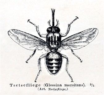
Tsetse are similar to other large flies, such as the housefly, but can be distinguished by various characteristics of their anatomy, two of which are easy to observe. Tsetse fold their wings completely when they are resting so that one wing rests directly on top of the other over their abdomens. Tsetse also have a long proboscis, which extends directly forward and is attached by a distinct bulb to the bottom of their heads.
Fossilized tsetse have been recovered from the Florissant Fossil Beds in Colorado, laid down some 34 million years ago. Twenty-three extant species of tsetse flies are known from Africa.
Tsetse fly giving birth
Etymology
The word tsetse means "fly" in Tswana, a Bantu language of southern Africa. Recently, tsetse without the fly has become more common in English, particularly in the scientific and development communities.
The word is pronounced tseh-tseh in the Sotho languages and is easily rendered in other African languages. During World War II, a de Havilland antisubmarine aircraft was known as the 'Tsetse' Mosquito.
Biology
The biology of tsetse is relatively well understood by Entomologists. They have been extensively studied because of their medical, veterinary, and economic importances, because the flies can be raised in a laboratory, and because they are relatively large, facilitating their analysis.
Morphology
Tsetse flies can be seen as independent individuals in two forms: as third-instar larvae, and as adults.
Tsetse first become separate from their mothers during the third larval instar, during which they have the typical appearance of maggots. However, this life stage is short, lasting at most a few hours, and is almost never observed outside of the laboratory.
Tsetse next develop a hard external case, the puparium, and become pupae—small, hard-shelled, oblongs with two distinctive, small, dark lobes at the tail (breathing) end. Tsetse pupae are under 1.0 cm long. Within the puparial shell, tsetse complete the last two larval instars and the pupal stage.
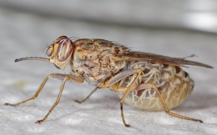
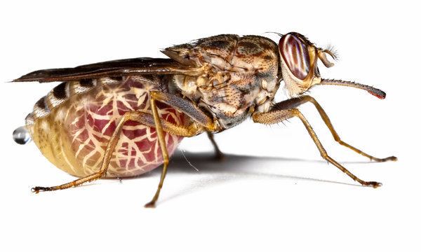
At the end of the pupal stage, tsetse emerge as adult flies. The adults are relatively large flies, with lengths of 0.5-1.5 cm, and have a recognizable shape or bauplan which makes them easy to distinguish from other flies. Tsetse have large heads, distinctly separated eyes, and unusual antennae. The thorax is quite large, while the abdomen is wide rather than elongated and shorter than the wings.
Four characteristics definitively separate adult tsetse from other kinds of flies:
Anatomy
Like all other insects, tsetse flies have an adult body comprising three visibly distinct parts: the head, the thorax and the abdomen.
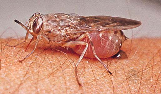
The head has large eyes, distinctly separated on each side, and a distinct, forward-pointing proboscis attached underneath by a large bulb. The thorax is large, made of three fused segments. Three pairs of legs are attached to the thorax, as are two wings and two halteres. The abdomen is short but wide and changes dramatically in volume during feeding.
The internal anatomy of tsetse is fairly typical of the insects. The crop is large enough to accommodate a huge increase in size during the bloodmeal since tsetse can take a bloodmeal equal in weight to themselves. The reproductive tract of adult females includes a uterus which can become large enough to hold the third-instar larva at the end of each pregnancy.
Most tsetse flies are physically very tough. Houseflies are easily killed with a flyswatter, but a great deal of effort is needed to crush a tsetse fly.
Life cycle
Tsetse have an unusual lifecycle which may be due to the richness of their food source. A female fertilizes only one egg at a time and retains each egg within her uterus to have the offspring develop internally during the first three larval stages, a method called adenotrophic viviparity. During this time, the female feeds the developing offspring with a milky substance secreted by a modified gland in the uterus. In the third larval stage, the tsetse larva leaves the uterus and begins its independent life. The newly independent tsetse larva crawls into the ground, and develops a hard outer shell called the puparial case, in which it completes its morphological transformation into an adult fly.
This lifestage has a variable duration, generally 20 to 30 days, and the larva must rely on stored resources during this time. The importance of the richness of blood to this development can be seen, since all tsetse development before it emerges from the puparial case as a full adult occurs without feeding, based only on nutritional resources provided by the female parent. The female must get enough energy for her needs, for the needs of her developing offspring, and for the stored resources which her offspring will require until it emerges as an adult.
Technically, these insects undergo the standard development process of insects, which consists of oocyte formation, ovulation, fertilization, development of the egg, three larval stages, a pupal stage, and the emergence and maturation of the adult.
Genetics
The genome of Glossina morsitans was sequenced in 2014.
Symbionts
Tsetse flies have three distinct symbionts. The primary symbiont is Wigglesworthia within the fly's bacteriocytes, the secondary symbiont is Sodalis intercellularly or intracellularly, and the third is some kind of Wolbachia.
The tsetse G. palpalis is also a vector and host of Hepatozoon petti, a parasitic sporozoan of the Nile crocodile.
Systematics
Tsetse are in the order Diptera, the true flies. They belong to the superfamily Hippoboscoidea, in which the tsetse's family, the Glossinidae, is one of four families of blood-feeding obligate parasites.
Up to 34 species and subspecies of tsetse flies are recognized, depending on the particular classification used.
All current classifications place all the tsetse species in a single genus named Glossina. Most classifications place this genus as the sole member of the family Glossinidae. The Glossinidae are generally placed within the superfamily Hippoboscoidea, which contains other hematophagous families.
Species
The tsetse genus is generally split into three groups of species based on a combination of distributional, behavioral, molecular and morphological characteristics. The genus includes:
Tsetse, hunger, and poverty
Tsetse flies are regarded as a major cause of rural poverty in sub-Saharan Africa because they prevent mixed farming. The land infested with tsetse flies is often cultivated by people using hoes rather than more efficient draught animals because nagana, the disease transmitted by tsetse, weakens and often kills these animals. Those cattle that survive produce little milk, pregnant cows often abort their calves, and manure is not available to fertilize the worn-out soils.
The disease nagana or African animal trypanosomiasis (AAT) causes gradual health decline in infected livestock, reduces milk and meat production, increases abortion rates, and animals eventually succumb to the disease (annual cattle deaths caused by trypanosomiasis are estimated at 3 million). This has an enormous impact on the livelihood of farmers who live in tsetse-infested areas, as infected animals cannot be used to plough the land, and keeping cattle is only feasible when the animals are kept under constant prophylactic treatment with trypanocidal drugs, often with associated problems of drug resistance, counterfeited drugs, and suboptimal dosage. The overall annual direct lost potential in livestock and crop production was estimated at US$4.5 billion.
The tsetse fly lives in nearly 10,000,000 square kilometres (4,000,000 sq mi) in sub-Saharan Africa and many parts of this large area is fertile land that is left uncultivated—a so-called green desert not used by humans and cattle. Most of the 37 countries infested with tsetse are poor, debt-ridden, and underdeveloped. Of the 39 tsetse-infested countries, 32 are low-income, food-deficit countries, 29 are least developed countries, and 30 are among the 40 most heavily indebted poor countries. Eradicating the tsetse and trypanosomiasis problem would allow rural Africans to reclaim areas of their continent and greatly increase food production. Only 45 million cattle, of 172 million present in sub-Saharan Africa, are kept in tsetse-infested areas, but are often forced into fragile ecosystems like highlands or the semiarid Sahel zone, which leads to overgrazing by animals and overuse of land by people for food production.
In addition to this direct impact, the presence of tsetse and trypanosomiasis discourages the use of more-productive exotic and cross-bred cattle, depresses the growth and affects the distribution of livestock populations, reduces the potential opportunities for livestock and crop production (mixed farming) through less draught power to cultivate land and less manure to fertilize (in an environment-friendly way) soils for better crop production, and affects human settlements (people tend to avoid areas with tsetse flies).
Tsetse flies transmit a similar disease to humans, called African trypanosomiasis - human African trypanosomiasis (HAT) or sleeping sickness. An estimated 70 million people in 20 countries are at different levels of risk and only 3-4 million people are covered by active surveillance. The DALY index (disability-adjusted life years), an indicator to quantify the burden of disease, includes the impact of both the duration of life lost due to premature death and the duration of life lived with a disability. The annual burden of sleeping sickness is estimated at 2 million DALYs. Since the disease tends to affect economically active adults, the total cost to a family with a patient is about 25% of a year’s income.
Trypanosomiasis
Tsetse are biological vectors of trypanosomes, meaning that in the process of feeding, they acquire and then transmit small, single-celled trypanosomes from infected vertebrate hosts to uninfected animals. Some tsetse-transmitted trypanosome species cause trypanosomiasis, an infectious disease. In humans, tsetse transmitted trypanosomiasis is called sleeping sickness. In animals, tsetse-vectored trypanosomiases include nagana, souma, and surra according to the animal infected and the trypanosome species involved. The usage is not strict and while nagana generally refers to the disease in cattle and horses it is commonly used for any of animal trypanosomiasis.
Trypanosomes are animal parasites, specifically protozoans of the genus Trypanosoma. These organisms are about the size of red blood cells. Different species of trypanosomes infect different hosts. They range widely in their effects on the vertebrate hosts. Some species, such as T. theileri, do not seem to cause any health problems except perhaps in animals that are already sick.
Some strains are much more virulent. Infected flies have an altered salivary composition which lowers feeding efficiency and consequently increases the feeding time, promoting trypanosome transmission to the vertebrate host. These trypanosomes are highly evolved and have developed a lifecycle that requires periods in both the vertebrate and tsetse hosts.
Tsetse transmit trypanosomes in two ways, mechanical and biological transmission.
The relative importance of these two modes of transmission for the propagation of tsetse-vectored trypanosomiases is not yet well understood. However, since the sexual phase of the trypanosome lifecycle occurs within the tsetse host, biological transmission is a required step in the lifecycle of the tsetse-vectored trypanosomes.
The cycle of biological transmission of trypanosomiasis involves two phases, one inside the tsetse host and the other inside the vertebrate host. Trypanosomes are not passed between a pregnant tsetse and her offspring, so all newly emerged tsetse adults are free of infection. An uninfected fly that feeds on an infected vertebrate animal may acquire trypanosomes in its proboscis or gut. These trypanosomes, depending on the species, may remain in place, move to a different part of the digestive tract, or migrate through the tsetse body into the salivary glands. When an infected tsetse bites a susceptible host, the fly may regurgitate part of a previous blood meal that contains trypanosomes, or may inject trypanosomes in its saliva. Inoculation must contain a minimum of 300 to 450 individual trypanosomes to be successful, and may contain up to 40,000 cells.
The trypanosomes are injected into vertebrate muscle tissue, but make their way, first into the lymphatic system, then into the bloodstream, and eventually into the brain. The disease causes the swelling of the lymph glands, emaciation of the body, and eventually leads to death. Uninfected tsetse may bite the infected animal prior to its death and acquire the disease, thereby closing the transmission cycle.
The tsetse-vectored trypanosomiases affect various vertebrate species including humans, antelopes, bovine cattle, camels, horses, sheep, goats, and pigs. These diseases are caused by several different trypanosome species that may also survive in wild animals such as crocodiles and monitor lizards. The diseases have different distributions across the African continent, so are transmitted by different species. This table summarizes this information:
Human trypanosomiasis
Human African trypanosomiasis, also called sleeping sickness, is caused by trypanosomes of the species Trypanosoma brucei. This disease is invariably fatal unless treated but can almost always be cured with current medicines, if the disease is diagnosed early enough.
Sleeping sickness begins with a tsetse bite leading to an inoculation in the subcutaneous tissue. The infection moves into the lymphatic system, leading to a characteristic swelling of the lymph glands called Winterbottom's sign. The infection progresses into the blood stream and eventually crosses into the central nervous system and invades the brain leading to extreme lethargy and eventually to death.
The species Trypanosoma brucei, which causes the disease, has often been subdivided into three subspecies that were identified based either on the vertebrate hosts which the strain could infect or on the virulence of the disease in humans. The trypanosomes infectious to animals and not to humans were named Trypanosoma brucei brucei. Strains that infected humans were divided into two subspecies based on their different virulences: Trypanosoma brucei gambiense was thought to have a slower onset and Trypanosoma brucei rhodesiense refers to strains with a more rapid, virulent onset. This characterization has always been problematic but was the best that could be done given the knowledge of the time and the tools available for identification. A recent molecular study using restriction fragment length polymorphism analysis suggests that the three subspecies are polyphyletic, so the elucidation of the strains of T. brucei infective to humans requires a more complex explanation. Procyclins are proteins developed in the surface coating of trypanosomes whilst in their tsetse fly vector.
Other forms of human trypanosomiasis also exist but are not transmitted by tsetse. The most notable is American trypanosomiasis, known as Chagas disease, which occurs in South America, caused by Trypanosoma cruzi, and transmitted by certain insects of the Reduviidae, members of the Hemiptera.
Animal trypanosomiasis
Animal trypanosomiasis, also called nagana when it occurs in bovine cattle or horses or sura when it occurs in domestic pigs, is caused by several trypanosome species. These diseases reduce the growth rate, milk productivity, and strength of farm animals, generally leading to the eventual death of the infected animals. Certain species of cattle are called trypanotolerant because they can survive and grow even when infected with trypanosomes although they also have lower productivity rates when infected.
The course of the disease in animals is similar to the course of sleeping sickness in humans.
Trypanosoma congolense and Trypanosoma vivax are the two most important species infecting bovine cattle in sub-Saharan Africa. Trypanosoma simiae causes a virulent disease in swine.
Other forms of animal trypanosomiasis are also known from other areas of the globe, caused by different species of trypanosomes and transmitted without the intervention of the tsetse fly.
The tsetse fly vector ranges mostly in the central part of Africa.
Control
The conquest of sleeping sickness and nagana would be of immense benefit to rural development and contribute to poverty alleviation and improved food security in sub-Saharan Africa. Human African trypanosomosis (HAT) and animal African trypanosomosis (AAT) are sufficiently important to make virtually any intervention against these diseases beneficial.
The disease can be managed by controlling the vector and thus reducing the incidence of the disease by disrupting the transmission cycle. Another tactic to manage the disease is to target the disease directly using surveillance and curative or prophylactic treatments to reduce the number of hosts that carry the disease.
Economic analysis indicates that the cost of managing trypanosomosis through the elimination of important populations of major tsetse vectors will be covered several times by the benefits of tsetse-free status. Area-wide interventions against the tsetse and trypanosomosis problem appear more efficient and profitable if sufficiently large areas, with high numbers of cattle, can be covered.
Vector control strategies can aim at either continuous suppression or eradication of target populations. Tsetse fly eradication programmes are complex and logistically demanding activities and usually involve the integration of different control tactics, such as trypanocidal drugs, impregnated treated targets (ITT), insecticide-treated cattle (ITC), aerial spraying (Sequential Aerosol Technique - SAT) and in some situations the release of sterile males (sterile insect technique – SIT). To ensure sustainability of the results, it is critical to apply the control tactics on an area-wide basis, i.e. targeting an entire tsetse population that is preferably genetically isolated.
Control techniques
Many techniques have reduced tsetse populations, with earlier, crude methods recently replaced by methods that are cheaper, more directed, and ecologically better.
Slaughter of wild animals
One early technique involved slaughtering all the wild animals tsetse fed on. For example, the island of Principe off the west coast of Africa was entirely cleared of feral pigs in the 1930s, which led to the extirpation of the fly. While the fly eventually re-invaded in the 1950s, the new population of tsetse was free from the disease.
Land clearing
Another early technique involved complete removal of brush and woody vegetation from an area. Tsetse tend to rest on the trunks of trees so removing woody vegetation made the area inhospitable to the flies. However, the technique was not widely used and has been abandoned. Preventing regrowth of woody vegetation requires continuous clearing efforts, which is only practical where large human populations are present. The clearing of woody vegetation has come to be seen as an environmental problem more than a benefit.
Pesticide campaigns
Pesticides have been used to control tsetse starting initially during the early part of the twentieth century in localized efforts using the inorganic metal based pesticides, expanding after the Second World War into massive aerial and ground based campaigns with organochlorine pesticides such as DDT applied as aerosol sprays at Ultra-Low Volume rates. Later, more targeted techniques used pour-on formulations in which advanced organic pesticides were applied directly to the backs of cattle.
Trapping
Tsetse populations can be monitored and effectively controlled using simple, inexpensive traps. These often use electric blue cloth, since this color attracts the flies. Early traps mimicked the form of cattle but this seems unnecessary and recent traps are simple sheets or have a biconical form. The traps can kill by channeling the flies into a collection chamber or by exposing the flies to insecticide sprayed on the cloth. Tsetse are also attracted to large dark colors like the hides of cow and buffaloes. Some scientists put forward the idea that zebra have stripes, not as a camouflage in long grass, but because the black and white bands tend to confuse tsetse and prevent attack.
The use of chemicals as attractants to lure tsetse to the traps has been studied extensively in the late 20th century, but this has mostly been of interest to scientists rather than as an economically reasonable solution. Attractants studied have been those tsetse might use to find food, like carbon dioxide, octenol, and acetone—which are given off in animals' breath and distributed downwind in an odor plume. Synthetic versions of these chemicals can create artificial odor plumes. A cheaper approach is to place cattle urine in a half gourd near the trap. For large trapping efforts, additional traps are generally cheaper than expensive artificial attractants.
A special trapping method is applied in Ethiopia, where the BioFarm Consortium (ICIPE, BioVision Foundation, BEA, Helvetas, DLCO-EA, Praxis Ethiopia) applies the traps in a sustainable agriculture and rural development context (SARD). The traps are just the entry point, followed by improved farming, human health and marketing inputs. This method is in the final stage of testing (as per 2006).
In the late 18th century, the Kotokoli Muslims of Togo held a special ritual in order for their child to have a prosperous life. This ritual consisted of mothers killing the tsetse flies and sprinkling them on horned melon. They would feed their children this delicacy. This ritual is still practiced today in some sub-Saharan tribes.
Sterile insect technique
The sterile insect technique (SIT) is a form of pest control that uses ionizing radiation (gamma ray or X ray) to sterilize male flies that are mass-produced in special rearing facilities. The sterile males are released systematically from the ground or by air in tsetse-infested areas, where they mate with wild females, which do not produce offspring. As a result, this technique can eventually eradicate populations of wild flies. SIT is among the most environmentally friendly control tactics available, and is usually applied as the final component of an integrated campaign.
The sustainable removal of the tsetse fly is in many cases the most cost-effective way of dealing with the T&T problem resulting in major economic benefits for subsistence farmers in rural areas. Insecticide-based methods are normally very ineffective in removing the last remnants of tsetse populations, while, on the contrary, sterile males are very effective in finding and mating the last remaining females. Therefore, the integration of the SIT as the last component of an area-wide integrated approach is essential in many situations to achieve complete eradication of the different tsetse populations, particularly in areas of more dense vegetation.
A project that was implemented from 1994 to 1997 on the Island of Unguja, Zanzibar (United Republic of Tanzania), demonstrated that, after suppression of the tsetse population with insecticides, SIT completely removed the Glossina austeni Newstead population from the Island (). The eradication of the tsetse fly from Unguja Island in 1997 was followed by the disappearance of the AAT which enabled farmers to integrate livestock keeping with cropping in areas where this had been impossible before. The increased livestock and crop productivity and the possibility of using animals for transport and traction significantly contributed to an increase in the quality of people’s lives (). A recent entomological survey (2015) jointly carried out by independent experts and the Department of Veterinary Services of Zanzibar has confirmed the tsetse-free status of the island, 18 years after eradication was declared.
In the Niayes region of Senegal, a coastal area close to Dakar, livestock keeping was difficult due to the presence of a population of Glossina palpalis gambiensis. Feasibility studies indicated that the fly population was confined to very fragmented habitats and a population genetics study indicated that the population was genetically isolated from the main tsetse belt in the south eastern part of Senegal. After completion of the feasibility studies (2006-2010), an area-wide integrated eradication campaign that included an SIT component was started in 2011, and by 2015, the Niayes region had become almost tsetse fly free.
The entire target area (Block 1, 2 and 3) has a total surface of 1000 km2, and the first block (northern part) can be considered free of tsetse, as intensive monitoring has failed to detect since 2012 a single wild tsetse fly. The prevalence of AAT has decreased from 40-50% before the project started to less than 10% to date in blocks 1 and 2. Although insecticides are being used for fly suppression, they are applied for short periods on traps, nets and livestock, and are not spread into the environment. After the suppression activities are completed, no more insecticide is applied in the area. The removal of trypanosomosis will eliminate the need for constant prophylactic treatments of the cattle with trypanocidal drugs, therefore reducing residues of these drugs in the dung, meat and milk.
The main beneficiaries of the project are the many small holder farmers, the larger commercial farms and the consumers of meat and milk. According to a socio-economic survey and benefit cost analysis, after eradication of the tsetse farmers will be able to replace their local breeds with improved breeds and increase their annual income by €2.8 million. In addition, it is expected that the number of cattle will be reduced by 45%, which will result in reduced environmental impacts.
Effect on societal development
In the literature of environmental determinism, the tsetse has been linked to difficulties during early state formation for areas where the fly is prevalent. In a 2012 article Dr. Marcella Alsan used population growth models, physiological data, and ethnographic data to examine pre-colonial agricultural practices and isolate the effects of the fly. Her results suggest that the tsetse decimated livestock populations, forcing early states to rely on slave labor to clear land for farming, and preventing farmers from taking advantage of natural animal fertilizers to increase crop production. Alsan argues that these long-term effects kept population density low and discouraged cooperation between small scale communities, thus preventing stronger nations from forming.
T. brucei sexual cycle
T. brucei are able to undergo meiotic sexual reproduction. Meiosis occurs within the salivary glands of the tsetse fly and is thought to be a normal part of development. The meiotic process results in production of haploid promastigote-like gametes. These gametes can interact with each other using their flagella, and then fuse.
Resistance of tsetse flies to trypanosome infection
Tsetse flies have an arsenal of immune defenses to resist each stage of the trypanosome infectious cycle, and thus are relatively refractory to trypanosome infection Among the host flies’ defenses is the production of hydrogen peroxide, a reactive oxygen species that damages DNA. These defenses limit the population of infected flies.