
Latin neostriatum TA A14.1.09.516 | NeuroLex ID Striatum FMA 77616 | |
 | ||
Part of Basal gangliaReward system Components Ventral striatumDorsal striatum | ||
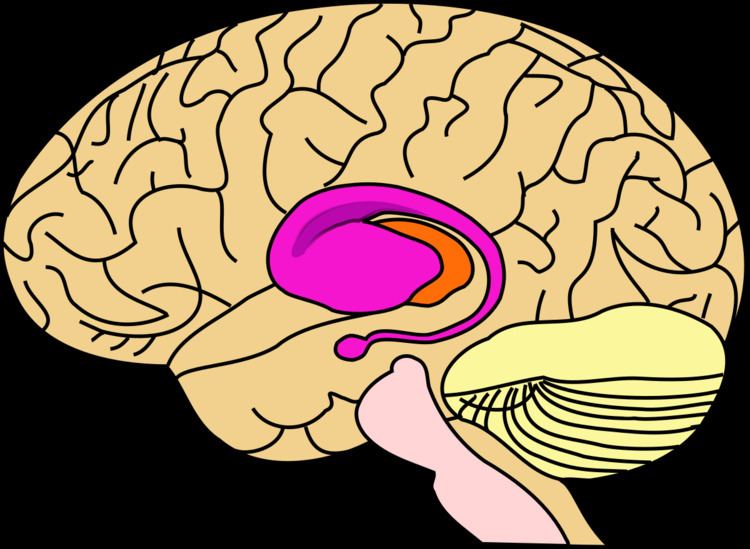
The striatum, also known as the neostriatum or striate nucleus, is one of the nuclei in the subcortical basal ganglia of the forebrain. The striatum is a critical component of the motor and reward systems. It receives both glutamatergic and dopaminergic inputs from different sources, and serves as the primary input to the rest of the basal ganglia nuclei.
Contents
- Structure
- Cell types
- Inputs
- Targets
- Function
- Parkinsons disease
- Addiction
- Bipolar disorder
- Dysfunction
- History
- Other animals
- References
In primates, the the striatum is divided into ventral and dorsal subdivisions, based upon function and connections. The ventral striatum consists of the nucleus accumbens and the olfactory tubercle. The dorsal striatum consists of the caudate nucleus and the putamen. A white matter tract called the internal capsule in the dorsal part separates the caudate nucleus and the putamen. The striatum functions to coordinate multiple aspects of cognition, including motor and action planning, decision-making, motivation, reinforcement, and reward perception. The term striatum is used to describe the striped (striated) appearance of the grey and white matter of this structure.
Sometimes the globus pallidus is included with the striatum when it is then known as the corpus striatum. The lentiform nucleus refers to the putamen of the dorsal striatum together with the globus pallidus.
Structure
The striatum is divided into ventral and dorsal subdivisions, based upon function and connections.
The ventral striatum is composed of the nucleus accumbens and the olfactory tubercle. The nucleus accumbens is made up of the nucleus accumbens core and nucleus accumbens shell, which differ by neuron populations. The olfactory tubercle receives input from the olfactory bulb but has not been shown to play a role in processing smell. In non-primate species, the islands of Calleja are included. The ventral striatum is associated with the limbic system and has been implicated as a vital part of the circuitry for decision making and reward-related behavior.
The dorsal striatum is composed of the caudate nucleus and the putamen. Staining can differentiate the dorsal striatum into compartments of striosomes and surrounding matrix; this is particularly evident on the components of acetylcholinesterase and calbindin.
Cell types
Types of cells in the striatum include:
The neurons of the striatum are not distributed evenly.
There are two regions of neurogenesis in the brain the subventricular zone and the dentate gyrus. Cells - neuroblasts, in the lateral ventricle adjacent to the striatum integrate in the striatum.
Inputs
The largest connection is from the cortex, in terms of cell axons. Many parts of the neocortex innervate the dorsal striatum. The cortical pyramidal neurons projecting to the striatum are located in layers II-VI, with the most dense projections come from layer V. They end mainly on the dendritic spines of the spiny neurons. They are glutamatergic, exciting striatal neurons.
The ventral striatum receives direct input from multiple regions in the cerebral cortex and limbic structures such as the amygdala, thalamus, and hippocampus, as well as the entorhinal cortex and the inferior temporal gyrus. Its primary input is to the basal ganglia system. Additionally, the mesolimbic pathway projects from the ventral tegmental area to the nucleus accumbens of the ventral striatum.
Another well-known afferent is the nigrostriatal connection arising from the neurons of the substantia nigra pars compacta. While cortical axons synapse mainly on spine heads of spiny neurons, nigral axons synapse mainly on spine shafts. In primates, the thalamostriatal afferent comes from the central median-parafascicular complex of the thalamus (see primate basal ganglia system). This afferent is glutamatergic. The participation of truly intralaminar neurons is much more limited. The striatum also receives afferents from other elements of the basal ganglia such as the subthalamic nucleus (glutamatergic) or the external globus pallidus (GABAergic).
Targets
The primary outputs of the ventral striatum project to the ventral pallidum, then the medial dorsal nucleus of the thalamus, which is part of the frontostriatal circuit. Additionally, the ventral striatum projects to the globus pallidus, and substantia nigra pars reticulata. Some of its other outputs include projections to the extended amygdala, lateral hypothalamus, and pedunculopontine nucleus.
Striatal outputs from both the dorsal and ventral components are primarily composed of medium spiny neurons (MSNs), a type of projection neuron, which have two primary phenotypes: "indirect" MSNs that express D2-type receptors and "direct" MSNs that express D1-type receptors.
The basal ganglia core is made up of the striatum along with the regions to which it projects directly, via the striato-pallidonigral bundle. The striato-pallidonigral bundle is a very dense bundle of sparsely myelinated axons, giving a whitish appearance. This projection comprises successively the external globus pallidus (GPe), the internal globus pallidus (GPi), the pars compacta of the substantia nigra (SNc), and the pars reticulata of substantia nigra (SNr). The neurons of this projection are inhibited by GABAergic synapses from the dorsal striatum. Among these targets, the GPe does not send axons outside the system. Others send axons to the superior colliculus. Two others comprise the output to the thalamus, forming two separate channels: one through the internal segment of the globus pallidus to the ventral oralis nuclei of the thalamus and from there to the cortical supplementary motor area and another through the substantia nigra to the ventral anterior nuclei of the thalamus and from there to the frontal cortex and the oculomotor cortex.
Function
The ventral striatum, and the nucleus accumbens in particular, primarily mediates reward cognition, reinforcement, and motivational salience, whereas the dorsal striatum primarily mediates cognition involving motor function, certain executive functions (e.g., inhibitory control), and stimulus-response learning; there is a small degree of overlap, as the dorsal striatum is also a component of the reward system that, along with the nucleus accumbens core, mediates the encoding of new motor programs associated with future reward acquisition (e.g., the conditioned motor response to a reward cue).
Metabotropic dopamine receptors are present both on spiny neurons and on cortical axon terminals. Second messenger cascades triggered by activation of these dopamine receptors can modulate pre- and postsynaptic function, both in the short term and in the long term. In humans, the striatum is activated by stimuli associated with reward, but also by aversive, novel, unexpected, or intense stimuli, and cues associated with such events. fMRI evidence suggests that the common property linking these stimuli, to which the striatum is reacting, is salience under the conditions of presentation. A number of other brain areas and circuits are also related to reward, such as frontal areas. Functional maps of the striatum reveal interactions with widely distributed regions of the cerebral cortex important to a diverse range of functions.
Parkinson's disease
Parkinson's disease results in loss of dopaminergic innervation to the dorsal striatum (and other basal ganglia) and a cascade of consequences. Atrophy of the striatum is also involved in Huntington's disease, choreas, choreoathetosis, and dyskinesias.
Addiction
Addiction, a disorder of the brain's reward system, arises through the overexpression of ΔFosB, a transcription factor, in the D1-type medium spiny neurons of the ventral striatum. ΔFosB is an inducible gene which is increasingly expressed in the nucleus accumbens as a result of repeatedly overdosing on an addictive drug or overexposure to other addictive stimuli.
Bipolar disorder
There is seen to be an association between striatal expression of variants of the PDE10A gene and some bipolar disorder I patients. Variants of other genes, DISCI and GNAS, have been associated with type II bipolar disorder.
Dysfunction
Dysfunction in the ventral striatum can lead to a variety of disorders, most notably, depression and obsessive-compulsive disorder. Because of its involvement in reward pathways, the ventral striatum has also been implicated in playing a critical role in addiction. It has been well established that the ventral striatum is strongly involved in mediating the reinforcing effects of drugs, especially stimulants, through dopaminergic stimulation.
History
In the seventeenth and eighteenth centuries, the term "corpus striatum" was used to designate many distinct, deep, infracortical elements of the hemisphere. In 1941, Cécile and Oskar Vogt simplified the nomenclature by proposing the term striatum for all elements built with striatal elements (see primate basal ganglia system): the caudate, the putamen, and the fundus striati, that ventral part linking the two preceding together ventrally to the inferior part of the internal capsule.
The term neostriatum was forged by comparative anatomists comparing the subcortical structures between vertebrates, because it was thought to be a phylogenetically newer section of the corpus striatum. The term is still used by some sources, including Medical Subject Headings.
Other animals
In birds the striatum is called the paleostriatum augmentatum.
In non-primate species, the islands of Calleja are included in the ventral striatum.