
Part of Temporal lobe NeuroNames hier-161 | Latin gyrus dentatus NeuroLex ID Dentate Gyrus | |
 | ||
Artery Posterior cerebralAnterior choroidal MeSH A08.186.211.577.405.200 | ||
The dentate gyrus is part of the hippocampus and/or hippocampal formation, as some texts include the latter structure in the former or vice versa. The dentate gyrus is thought to contribute to the formation of new episodic memories, the spontaneous exploration of novel environments, and other functions. It is notable as being one of a select few brain structures currently known to have high rates of neurogenesis in adult rats (other sites include the olfactory bulb and cerebellum).
Contents
Granule cells within the molecular layer of the dentate gyrus receive the hippocampal formation's major excitatory input from the cortex. This input is primarily made up of signals from layer II of the entorhinal cortex and is the first connection of the trisynaptic loop, the hippocampal circuit.
Structure
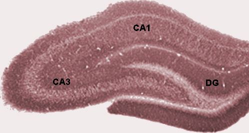
The dentate gyrus is composed of unidirectional projections dispersed towards CA3 pyramidal cells of the hippocampus. It consists of three layers of neurons: molecular, granular, and polymorphic. The granular layer is most prominent and contains granule cells that project to the CA3 subfield of the hippocampus, as previously mentioned. These granule cells project mostly to interneurons, but also to pyramidal cells and are the principal excitatory neurons of the dentate gyrus. The major input to the dentate gyrus (the so-called perforant pathway) is from layer 2 of the entorhinal cortex, and the dentate gyrus receives no direct inputs from other cortical structures. The perforant pathway is divided into the medial and lateral perforant paths, generated, respectively, at the medial and lateral portions of the entorhinal cortex. The medial perforant path synapses onto the proximal dendritic area of the granule cells, whereas the lateral perforant path does so onto the distal dendrites of these same cells. Most lateral views of the dentate gyrus may appear to suggest a structure consisting of just one entity, but medial movement may provide evidence of the ventral and dorsal parts of the dentate gyrus.
Development
The granule cells in the dentate gyrus are distinguished by their late time of formation during brain development. In rats, approximately 85% of the granule cells are generated after birth. In humans, it is estimated that granule cells begin to be generated during gestation weeks 10.5 to 11, and continue being generated during the second and third trimesters, after birth and all the way into adulthood. The germinal sources of granule cells and their migration pathways have been studied during rat brain development. The oldest granule cells are generated in a specific region of the hippocampal neuroepithelium and migrate into the primordial dentate gyrus around embryonic days (E) 17/18, and then settle as the outermost cells in the forming granular layer. Next, dentate precursor cells move out of this same area of the hippocampal neuroepithelium and, retaining their mitotic capacity, invade the hilus (core) of the forming dentate gyrus. This dispersed germinal matrix is the source of granule cells from that point on. The newly generated granule cells accumulate under the older cells that began to settle in the granular layer. As more granule cells are produced, the layer thickens and the cells are stacked up according to age - the oldest being the most superficial and the youngest being deeper. The granule cell precursors remain in a subgranular zone that becomes progressively thinner as the dentate gyrus grows, but these precursor cells are retained in adult rats. These sparsely scattered cells constantly generate granule cell neurons, which add to the total population. There are a variety of other differences in the rat, monkey and human dentate gyrus. The granule cells only have apical dendrites in the rat. But in the monkey and human, many granule cells also have basal dendrites.
Function
The dentate gyrus is thought to contribute to the formation of memories, and to play a role in depression.
Memory
The dentate gyrus is one of the few regions of the adult brain where neurogenesis (i.e., the birth of new neurons) takes place. Neurogenesis is thought to play a role in the formation of new memories. New memories could preferentially use newly formed dentate gyrus cells, providing a potential mechanism for distinguishing multiple instances of similar events or multiple visits to the same location. This increased neurogenesis is associated with improved spatial memory, as seen through performance in a maze.
Stress and depression
The dentate gyrus may also have a functional role in stress and depression. For instance, neurogenesis has been found to increase in response to chronic treatment with antidepressants. The physiological effects of stress, often characterized by release of glucocorticoids such as cortisol, as well as activation of the sympathetic division of the autonomic nervous system, have been shown to inhibit the process of neurogenesis in primates. Both endogenous and exogenous glucocorticoids are known to cause psychosis and depression, implying that neurogenesis in the dentate gyrus may play an important role in modulating symptoms of stress and depression.
Other
Some evidence suggests neurogenesis in the dentate gyrus increases in response to aerobic exercise. Several experiments have shown neurogenesis (the development of nerve tissues) often increases in the dentate gyrus of adult rodents when they are exposed to an enriched environment. The dentate gyrus is also known to serve as a pre-processing unit. When information enters, it is known to separate very similar information into distinct and unique details. This prepares the relevant data for storage in the hippocampal CA3 section.
Spatial behavior
Studies have shown that after destroying about 90% of their dentate gyrus (dg) cells, rats had extreme difficulty in maneuvering through a maze they had been through, prior to the lesion being made. When being tested a number of times to see whether they could learn a maze, the results showed that the rats did not improve at all, indicating that their working memories were severely impaired. Rats had trouble with place strategies because they could not consolidate learned information about a maze into their working memory, and, thus, could not remember it when maneuvering through the same maze in a later trial. Every time a rat entered the maze, the rat behaved as if it was seeing the maze for the first time.
Blood sugar
Studies by researchers at Columbia University Medical Center indicate poor glucose control can lead to deleterious effects on the dentate gyrus.