
Symbol PVALB HUGO 9704 RefSeq NM_002854 | Entrez 5816 OMIM 168890 UniProt P20472 | |
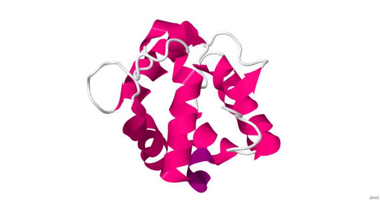 | ||
Parvalbumin is a calcium-binding albumin protein with low molecular weight (typically 9-11 kDa).
Contents
It has three EF hand motifs and is structurally related to calmodulin and troponin C. Parvalbumin is localised in fast-contracting muscles, where its levels are highest, as well as in the brain and some endocrine tissues.
Parvalbumin is a small, stable protein containing EF-hand type calcium binding sites. It is involved in calcium signaling. Typically, this protein is broken into three domains, domains AB, CD and EF, each individually containing a helix-loop-helix motif. The AB domain houses a two amino-acid deletion in the loop region, whereas domains CD and EF contain the N-terminal and C-terminal, respectively.
Calcium binding proteins like parvalbumin play a role in many physiological processes, namely cell-cycle regulation, second messenger production, muscle contraction, organization of microtubules and phototransduction. Alterations in the function of parvalbumin-expressing neurons have been implicated in various areas of clinical interest such as Alzheimer's disease, age-related cognitive defects and some forms of cancer.
Location and function
Parvalbumin (PV) is present in GABAergic interneurons in the nervous system, especially the reticular thalamus, and expressed predominantly by chandelier and basket cells in the cortex. In the cerebellum, PV is expressed in Purkinje cells and molecular layer interneurons. In the hippocampus, PV+ interneurons are subdivided into basket, axo-axonic, bistratified, and oriens-lacunosum moleculare (O-LM) cells, each subtype targeting distinct domains of pyramidal cells.
PV interneurons' connections are mostly perisomatic (around the cell body of neurons). Most of the PV interneurons are fast-spiking. They are also thought to give rise to gamma waves recorded in EEG.
PV-expressing interneurons represent approximately 25% of GABAergic cells in the primate DLPFC. Other calcium-binding protein markers are calretinin (most abundant subtype in DLPFC, about 50%) and calbindin. Interneurons are also divided into subgroups by the expression of neuropeptides such as somatostatin, neuropeptide Y, cholecystokinin.
Role in pathology
Decreased PV and GAD67 expression was found in PV+ GABAergic interneurons in schizophrenia. PV has been identified as an allergen causing fish allergy.