
Latin substantia alba TA A14.1.00.009 | Dorlands/Elsevier White matter FMA 83929 | |
 | ||
White matter refers to areas of the central nervous system that are mainly made up of myelinated axons, also called tracts. Long thought to be passive tissue, white matter actively affects learning and brain functions, modulating the distribution of action potentials, acting as a relay and coordinating communication between different brain regions.
Contents
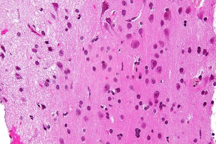
White matter is named for its relatively light appearance resulting from the lipid content of myelin. However, the tissue of the freshly cut brain appears pinkish white to the naked eye because myelin is composed largely of lipid tissue veined with capillaries. Its white color in prepared specimens is due to its usual preservation in formaldehyde.
White matter
White matter is composed of bundles of myelinated nerve cell projections (or axons), which connect various gray matter areas (the locations of nerve cell bodies) of the brain to each other, and carry nerve impulses between neurons. Myelin acts as an insulator, which allows electrical signals to jump, rather than coursing through the axon, increasing the speed of transmission of all nerve signals.
The total number of long range fibers within a cerebral hemisphere is 2% of the total number of cortico-cortical fibers (across cortical areas) and is roughly the same number as those that communicate between the two hemispheres in the brain's largest white tissue structure, the Corpus callosum. Schüz and Braitenberg note "As a rough rule, the number of fibres of a certain range of lengths is inversely proportional to their length."
White matter in nonelderly adults is 1.7–3.6% blood.
Grey matter
The other main component of the brain is grey matter (actually pinkish tan due to blood capillaries), which is composed of neurons. The substantia nigra is a third colored component found in the brain that appears darker due to higher levels of melanin in dopaminergic neurons than its nearby areas. Note that white matter can sometimes appear darker than grey matter on a microscope slide because of the type of stain used. Cerebral- and spinal white matter do not contain dendrites, neural cell bodies, or shorter axons, which can only be found in grey matter.
Location
White matter forms the bulk of the deep parts of the brain and the superficial parts of the spinal cord. Aggregates of gray matter such as the basal ganglia (caudate nucleus, putamen, globus pallidus, subthalamic nucleus, nucleus accumbens) and brain stem nuclei (red nucleus, substantia nigra, cranial nerve nuclei) are spread within the cerebral white matter.
The cerebellum is structured in a similar manner as the cerebrum, with a superficial mantle of cerebellar cortex, deep cerebellar white matter (called the "arbor vitae") and aggregates of grey matter surrounded by deep cerebellar white matter (dentate nucleus, globose nucleus, emboliform nucleus, and fastigial nucleus). The fluid-filled cerebral ventricles (lateral ventricles, third ventricle, cerebral aqueduct, fourth ventricle) are also located deep within the cerebral white matter.
Myelinated axon length
Men have more white matter than females both in volume and in length of myelinated axons. At the age of 20, the total length of myelinated fibers in males is 176,000 km while that of a female is 149,000 km. There is a decline in total length with age of about 10% each decade such that a man at 80 years of age has 97,200 km and a female 82,000 km. Most of this reduction is due to the loss of thinner fibers.
Function
White matter is the tissue through which messages pass between different areas of gray matter within the central nervous system. The white matter is white because of the fatty substance (myelin) that surrounds the nerve fibers (axons). This myelin is found in almost all long nerve fibers, and acts as an electrical insulation. This is important because it allows the messages to pass quickly from place to place.
Unlike gray matter, which peaks in development in a person's twenties, the white matter continues to develop, and peaks in middle age. This claim has been disputed in recent years, however.
Clinical significance
Multiple sclerosis (MS) is one of the most common diseases which affect white matter. In MS lesions, the myelin shield around the axons has been destroyed by inflammation.
Alcohol use disorders are associated with decrease in white matter volume. Animal studies suggest that alcohol may cause loss of white matter by damaging oligodendrocytes, the glial cell responsible for maintaining myelin.
Changes in white matter known as amyloid plaques are associated with Alzheimer's disease and other neurodegenerative diseases. White matter injuries ("axonal shearing") may be reversible, while gray matter regeneration is less likely. Other changes that commonly occur with age include the development of leukoaraiosis, which is a rarefaction of the white matter that can be caused by a variety of conditions, including loss of myelin, axonal loss, and a breakdown of the blood–brain barrier.
Imaging
The study of white matter has been advanced with the neuroimaging technique called diffusion tensor imaging where magnetic resonance imaging (MRI) brain scanners are used. As of 2007, more than 700 publications have been published on the subject.
A 2009 paper by Jan Scholz and colleagues used diffusion tensor imaging (DTI) to demonstrate changes in white matter volume as a result of learning a new motor task (e.g. juggling). The study is important as the first paper to correlate motor learning with white matter changes. Previously, many researchers had considered this type of learning to be exclusively mediated by dendrites, which are not present in white matter. The authors suggest that electrical activity in axons may regulate myelination in axons. Or, gross changes in the diameter or packing density of the axon might cause the change. A more recent DTI study by Sampaio-Baptista and colleagues reported changes in white matter with motor learning along with increases in myelination.