
Location Basal ganglia Neurotransmitter GABA | Morphology Spiny neuron | |
 | ||
Function Inhibitory projection neuron Presynaptic connections Dopaminergic: VTA, SNcGlutamatergic: PFC, hippocampus, amygdala, thalamus, other Postsynaptic connections Other basal ganglia structures | ||
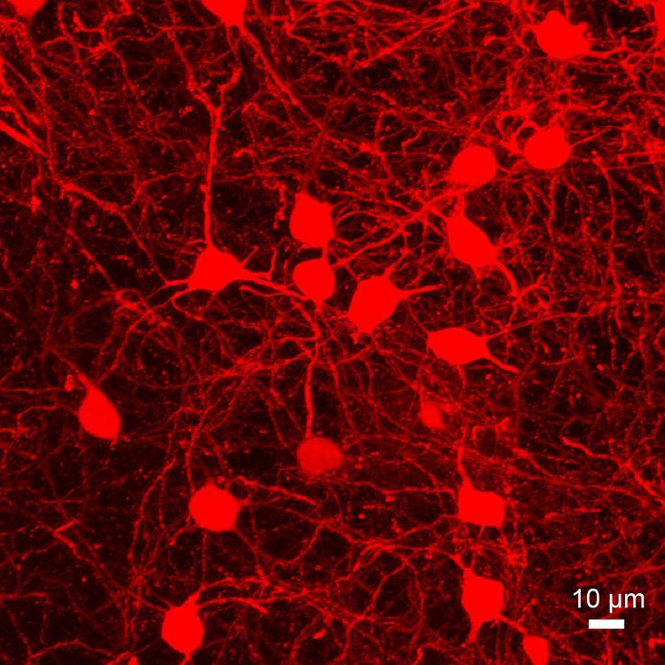
Medium spiny neurons (MSNs), also known as spiny projection neurons, are a special type of GABAergic inhibitory cell representing 95% of neurons within the human striatum, a structure located in the basal ganglia. Medium spiny neurons have two primary phenotypes (i.e., characteristic types): D1-type MSNs of the direct pathway and D2-type MSNs of the indirect pathway. Most striatal MSNs contain only D1-type or D2-type dopamine receptors, but a subpopulation of MSNs exhibit both phenotypes.
Contents
Direct pathway MSNs excite their ultimate basal ganglia output structure (e.g., the thalamus) and promote associated behaviors; these neurons express D1-type dopamine receptors, adenosine A1 receptors, dynorphin peptides, and substance P peptides. Indirect pathway MSNs inhibit their output structure and in turn inhibit associated behaviors; these neurons express D2-type dopamine receptors, adenosine A2A receptors (A2A), DRD2–A2A heterotetramers, and enkephalin. Both types express glutamate receptors (NMDAR and AMPAR) and CB1 receptors. A subpopulation of MSNs contain both D1-type and D2-type receptors, with approximately 40% of striatal MSNs expressing both DRD1 and DRD2 mRNA. In the nucleus accumbens (NAcc), these mixed-type MSNs that contain both D1-type and D2-type receptors are mostly contained in the NAcc shell.
The dorsal striatal MSNs play a key role in initiating and controlling movements of the body, limbs, and eyes. The ventral striatal MSNs play a key role in motivation, reward, reinforcement, and aversion. Dorsal and ventral medium spiny neuron subtypes (i.e., direct D1-type and indirect D2-type) are identical phenotypes, but their output connections differ.
Appearance and location
The medium spiny neurons are medium-sized neurons (~15 microns in diameter, ~12-13 microns in the mouse) with large and extensive dendritic trees (~500 microns in diameter). Striatal direct pathway MSNs (dMSNs) project directly to the globus pallidus internal (GPi) and substantia nigra pars reticulata (SNr) whereas striatal indirect pathway MSNs (iMSNs) ultimately project to these two structures via an intermediate connection to the globus pallidus external (GPe) and ventral pallidum (VP). The GPe and VP send a GABAergic projection to the subthalamic nucleus, which then sends glutamatergic projections to the GPi and SNr. Both the GPi and SNr send inhibitory projections to nuclei within the thalamus.
Function
MSNs are inhibitory GABAergic neurons, but the effect of direct MSNs (dMSNs) and indirect MSNs (iMSNs) on their ultimate output structures differs: dMSNs excite, while iMSNs inhibit, their basal ganglia output structures (e.g., the thalamus). Within the basal ganglia, there are several complex circuits of neuronal loops all of which include medium spiny neurons.
The cortical, thalamic, and brain-stem inputs that arrive at the medium spiny neurons show a vast divergence in that each incoming axon forms contacts with many spiny neurons and each spiny neuron receives a vast amount of input from different incoming axons. Since these inputs are glutamatergic they exhibit an excitatory influence on the inhibitory medium spiny neurons.
There are also interneurons in the striatum which regulate the excitability of the medium spiny neurons. The synaptic connections between a particular GABAergic interneuron, the parvalbumin expressing fast-spiking interneuron, and spiny neurons are close to the spiny neurons' soma, or cell body. Recall that excitatory postsynaptic potentials caused by glutamatergic inputs at the dendrites of the spiny neurons only cause an action potential when the depolarization wave is strong enough upon entering the cell soma. Since the fast-spiking interneurons influence is located so closely to this critical gate between the dendrites and the soma, they can readily regulate the generation of an action potential. Additionally, other types of GABAergic interneurons make connections with the spiny neurons. These include interneurons that express tyrosine hydroxylase and neuropeptide Y.
Direct pathway
The direct pathway within the basal ganglia makes excitatory inputs coming from e.g. the cortex cause a net excitation of upper motor neurons in the motor areas of the cortex. In the direct pathway, the medium spiny neurons project to the internal division of the globus pallidus which in turn sends axons to the substantia nigra pars reticulata (SNr) and the ventroanterior and ventrolateral thalamus (VTh). The SNr projects to the deep layer of the superior colliculus thus controlling fast eye movements (saccades). The VTh projects to upper motor neurons in the primary motor cortex (precentral gyrus).
Neurons in the globus pallidus are also inhibitory, thus inhibiting the excitatory neurons in the SNr and VTh. But in contrast to the medium spiny neurons, globus pallidus neurons are tonically active when not activated. Thus in the absence of cortical stimulation, SNr and VTh neurons are tonically inhibited thus preventing involuntary spontaneous movements.
Once the medium spiny neurons receive sufficient excitatory cortical input, they are excited and fire a burst of inhibitory action potentials to globus pallidus neurons. These tonically active neurons are then inhibited, causing their inhibitory influence on SNr and VTh to decline. Thus SNr and VTh neurons are disinhibited resulting in net excitement causing them to activate upper motor neurons commanding a movement. Cortical activation of the basal ganglia thus eventually results in excitement (disinhibition) of motor neurons causing movement to take place.
Indirect pathway
In the indirect pathway, excitation (e.g. cortical input to the basal ganglia) results in net inhibition of upper motor neurons. In this pathway the medium spiny neurons in the striatum project to the external segment of the globus pallidus. These neurons in turn project to the internal segment of the globus pallidus and to the subthalamic nuclei which forms a feedback loop to the internal globus pallidus.
Cortical excitement of medium spiny neurons causes them to inhibit external globus pallidus neurons. These tonically inhibiting neurons thus decrease their inhibitory influence on the internal globus pallidus and the subthalamic nuclei.
Globus pallidus neurons tonically inhibit VTh and SNr neurons. Since the inhibitory influence from the external globus pallidus is now reduced, these neurons show stronger activity thus increasing their inhibition of SNr and VTh neurons.
The projections of the external globus pallidus to the subthalamic nuclei causes these neurons to increase their firing rate, since the globus pallidus neurons are inhibited by medium spiny neurons. The subthalamic nuclei have excitatory projections to the internal globus pallidus thus causing the internal globus pallidus neurons to increase their inhibititory influence on SNr and VTh.
Eventually excitatory inputs from the cortex results in net inhibition of upper motor neurons thus preventing them from initiating a movement.