
Specialty medical genetics DiseasesDB 11252 | ICD-10 Q60.6 MedlinePlus 001268 | |
 | ||
ICD-9-CM 753.0, 658.0, 761.2 (for oligohydramnios) | ||
Potter sequence is the atypical physical appearance of a baby due to oligohydramnios experienced when in the uterus. Oligohydramnios is the decrease in amniotic fluid volume sufficient to cause deformations in morphogenesis of the fetus.
Contents
Oligohydramnios is the cause of Potter sequence but there are many things that can lead to oligohydramnios. It can be caused by renal diseases such as bilateral renal agenesis (BRA), atresia of the ureter or urethra causing obstruction of the urinary tract, polycystic or multicystic kidney diseases, renal hypoplasia, amniotic rupture, toxemia, or uteroplacental insufficiency from maternal hypertension.
Potter's sequence is known in the medical field as clubbed feet, pulmonary hypoplasia and cranial anomalies related to the oligohydramnios.
The term Potter sequence was initially intended to only refer to cases caused by BRA, however, it is now commonly used by many clinicians and researchers to refer to any case that presents with oligohydramnios or anhydramnios regardless of the source of the loss of amniotic fluid.
Signs and symptoms
The failure of the metanephros to develop in cases of BRA and some cases involving unilateral renal agenesis (URA) is due primarily to the failure of the mesonephric duct to produce a ureteric bud capable of inducing the metanephric mesenchyme. The failed induction will thereby cause the subsequent degeneration of the metanephros by apoptosis and other mechanisms. The mesonephric duct(s) of the agenic kidney(s) will also degenerates and fail to connect with the bladder. Therefore, the means by which the fetus produces urine and transports it to the bladder for excretion into the amniotic sac has been severely compromised (in the cases of URA), or completely eliminated (in the cases of BRA). The decreased volume of amniotic fluid causes the growing fetus to become compressed by the mother's uterus. This compression can cause many physical deformities of the fetus, most common of which is Potter facies. Lower extremity anomalies are frequent in these cases, which often presents with clubbed feet and/or bowing of the legs. Sirenomelia, or "Mermaid syndrome" (which occurs approximately in 1:45,000 births) can also present. In fact, nearly all reported cases of sirenomelia also be present with BRA.
Other anomalies of the classic Potter sequence infant include a parrot beak nose, redundant skin, and the most common characteristic of infants with BRA which is a skin fold of tissue extending from the medial canthus across the cheek. The ears are slightly low and pressed against the head making them appear large. The adrenal glands often appear as small oval discs pressed against the posterior abdomen due to the absence of upward renal pressure. The bladder is often small, nondistensible and may be filled with a minute amount of fluid. In males the vas deferens and seminal vesicles may be absent, while in females the uterus and upper vagina may be absent. Other abnormalities include anal atresia, absence of the rectum and sigmoid colon, esophageal and duodenal atresia, and a single umbilical artery. Presence of a diaphragmatic hernia is also common in these fetuses/infants. Additionally, the alveolar sacs of the lungs fail to properly develop as a result of the reduced volume of amniotic fluid. Labor is often induced between 22 and 36 weeks of gestation (however, some of these pregnancies may go to term) and unaborted infants typically survive for only a few minutes to a few hours. These infants will eventually die as either a result of pulmonary hypoplasia or renal failure.
Genetics
While genetic research has linked certain genetic mutations to be the cause of ARPKD, ADPKD and possibly MRD, to date no genetic mutation or chromosomal anomaly has been linked to be the cause of BRA. Chromosomal anomalies have been associated with BRA in certain cases (chromosomes 1, 2, 5 and 21), but these anomalies were not inherited and have not been observed in subsequent cases. Additionally, neither extreme substance abuse or environmental factors (high power line, mercury, etc.) have been reported to be linked to an increased incidence of BRA or other cause of Potter sequence. BRA and other causes of oligohydramnios sequence have been linked to a number of other problems, to include Down syndrome, Kallmann syndrome, branchio-oto-renal syndrome and others.
The high-risk obstetrician or genetic counselor may ask for a blood sample from the fetus or will perform an amniocentesis. These samples are used to perform several tests, one of which may be to check for the proper number of chromosomes, called a karyotype, of the fetus. Some birth defects are known to be associated with missing a chromosome, having an extra chromosome, such as in Down syndrome, as well as by having a part of one chromosome break off and relocate to a portion of another chromosome (called a translocation). However, on each of the 23 pairs of chromosomes are thousands of different genes. While chromosomes are easy to visualize under a microscope and count, the genes on them are not. Genes are very small pieces of DNA when compared to the chromosomes they reside on. A gene contains a code for a protein and if the gene is mutated (different from normal) the protein that is made from it may not function properly - if at all. Unfortunately, genetic abnormalities could still exist despite having normal chromosomes. The only way to determine genetically inherited mutations in the infant is to perform a genome scan of the mother, father, affected infant and any unaffected siblings of the affected fetus. These analyses will reveal what genetic mutations are present in the affected infant, and by comparing these results to the surviving siblings and parents, it can be determined which mutations were inherited or were not.
Classic form
Classic Potter sequence occurs when the developing fetus has bilateral renal agenesis, which also presents with agenesis of the ureters. BRA has been estimated to occur at a frequency of approximately 1:4000 to 1:8000 fetuses and neonates. However, recent analysis has estimated that the condition may occur at a much greater frequency. The condition has been reported to occur twice as commonly in males as in females, suggesting that certain genes of the Y chromosome may act as modifiers. However, no candidate genes on the Y chromosome have yet been identified.
BRA appears to have a predominantly genetic etiology and many cases represent the most severe manifestation of an autosomal dominant condition with incomplete penetrance and variable expressivity. There are several genetic pathways that could result in this condition. To date, few of these pathways or candidate genes have been considered or analyzed regarding BRA. The majority of possible candidate genetic pathways are autosomal recessive in nature and do not coincide with the frequency or penetrance at which BRA occurs in the human population. Additionally, candidate genetic pathways would be expected to involve genes expressed in the developing urogenital system (UGS). Often, these same genes and/or pathways of interacting genes are also expressed in the developing UGS as well as the central nervous system (CNS), gut, lung, limbs, and eyes.
Fetal urine
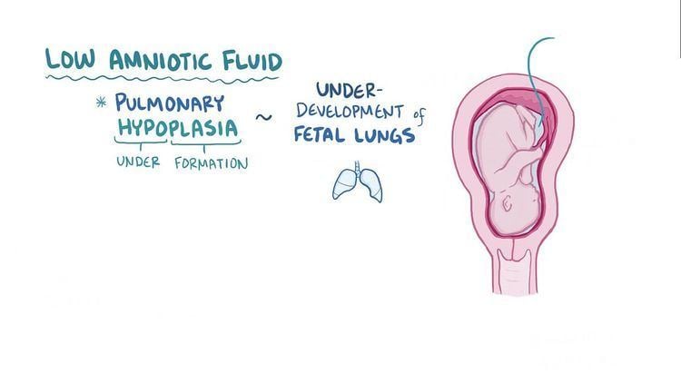
Development of the mature kidney begins between weeks 5 and 7 of gestation. Fetal urine production begins in early gestation and comprises the majority of the amniotic fluid in the second and third trimesters of pregnancy. The fetus continuously swallows amniotic fluid, which is reabsorbed by the gastrointestinal tract and then reintroduced into the amniotic cavity by the kidneys via urination. Oligohydramnios occurs if the volume of amniotic fluid is less than normal for the corresponding period of gestation. The fetal urine is critical to the proper development of the lungs by aiding in the expansion of the airways - alveoli, by means of hydrodynamic pressure and by also supplying proline which is a critical amino acid for lung development. Alveoli are the small sacs in the lungs that exchange oxygen with the blood. If the alveoli, and thereby the lungs, are underdeveloped at the time of birth the infant will not be able to breathe air properly and will go into respiratory distress shortly after birth due to pulmonary hypoplasia (underdeveloped lungs). This is the primary cause of death to Potter sequence infants secondary to renal failure. The fetal urine also serves to cushion the fetus from being compressed by the mother's uterus as it grows.
Types
Since its initial characterization, Potter sequence has been defined into five distinct subclassifications. There are those in the medical and research fields that use the term Potter sequence to specifically refer to only cases of BRA, while other groups use the term to loosely refer to all instances of oligohydramnios and anhydramnios regardless of the specific cause. The assignment of nomenclature to the various causes (types) was employed in order to help clarify these discrepancies, but these subclassifications and nomenclature system have not caught on in the medical and research communities.
Prognosis
In previously recorded medical and research history, BRA has proved to be lethal in all cases of singleton births. Various other forms of the sequence are, or are near, lethal in 100% of the cases.
The first child to survive Bilateral Renal Agenesis (BRA), Abigail Rose Herrera Beutler, was born on July 2013 to US Congresswoman Jaime Herrera Beutler. A few weeks before she was born, Dr. Jessica Bienstock, a professor of maternal-fetal medicine at John's Hopkins Hospital, administered a series of saline solution injections into the mother's womb to help the baby's lungs to develop. After Abigail was born, the procedure was considered a success. The infant did not need artificial respiration and could breathe on her own. Her parents kept her on kidney dialysis at home until old enough for a kidney transplant. On February 8, 2016, at the age of two, Abigail received a kidney from her father at the Lucile Packard Children's Hospital Stanford in California.
History
Bilateral renal agenesis (BRA) was first recognized as a defect of human fetal development in 1671 by Wolfstrigel.
In 1946, Edith Potter (1901 - 1993) described a series of 20 cases with absent kidneys, noting the characteristic appearance of the head and lungs. Up until this time, the condition itself was considered to be extremely rare. However, in part to Potter's work, it has come to light that the condition presents far more frequently than previously reported. Potter analyzed approximately 5000 autopsy cases performed on fetuses and newborn infants over a period of ten years and found that 20 of these infants presented with BRA, all of which had distinctive facial characteristics which did not appear to them to have any specific embryologic correlation with the renal anomaly. It was only much later when she and others attributed the multiple congenital deformities, including the features of Potter's facies and also pulmonary hypoplasia, to have been caused by the prolonged severe lack of amniotic fluid. These facial characteristics have subsequently been termed as being known as Potter facies. From her analysis, she was able to deduce the sequence of events that leads to what is now known as Potter sequence.
Potter went on to become a pioneer in the field of human renal development and her contributions are still employed and appreciated by clinicians and researchers to this day.
Terminology
Potter syndrome is not technically a syndrome as it does not collectively present with the same telltale characteristics and symptoms in each and every case. It is more accurately described as a "sequence" or chain of events that may have different beginnings (absent kidneys, cystic kidneys, obstructed ureters or other causes), but which all end with the same conclusion (absent or reduced volume of amniotic fluid). This is why Potter syndrome is often called Potter sequence or oligohydramnios sequence by some clinicians and researchers. The term Potter syndrome is most frequently associated with the condition of oligohydramnios sequence regardless of the root cause of the absence or reduced volume of amniotic fluid. However, as noted in this article, the term Potter syndrome was initially coined in order to refer to fetuses and infants with BRA. It was not until later that the term became more encompassing as it was noted that other causes of failed fetal urine production also resulted in similar physical characteristics and prognoses of the fetuses and infants with BRA (that which Potter originally described in 1946). Since then, the term Potter syndrome has become a misnomer and experts have attempted not to eliminate the terminology, but to modify it in a way so as to be able to determine the different root causes by creating a nomenclature system. However, this classification system has not caught on in the clinical and research fields.