
Latin paries abdominalis | FMA 299989 | |
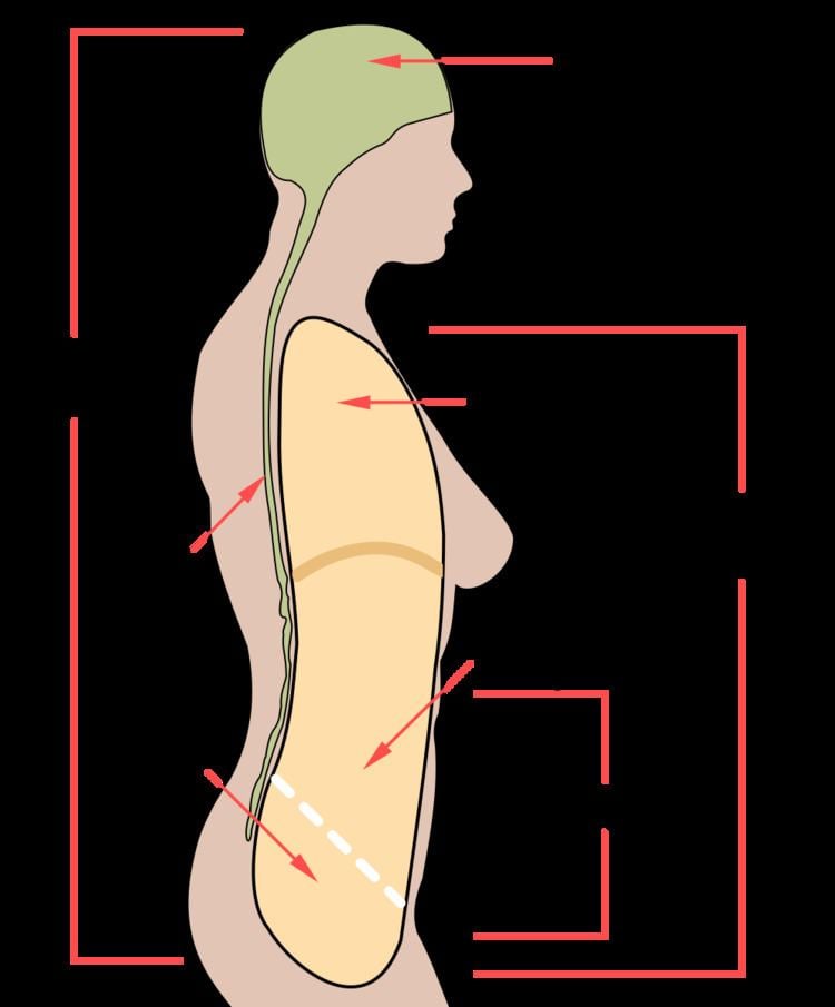 | ||
In anatomy, the abdominal wall represents the boundaries of the abdominal cavity. The abdominal wall is split into the posterior (back), lateral (sides) and anterior (front) walls.
Contents
There is a common set of layers covering and forming all the walls: the deepest being the parietal peritoneum, the extraperitoneal fat, the transversalis fascia, the internal and external oblique and transversus abdominis aponeuroses, and a layer of fascia, which has different names according to what it covers (e.g., transversalis, psoas fascia).
In medical vernacular, the abdominal wall most commonly refers to the layers composing the anterior abdominal wall which, in addition to the layers mentioned above, includes the three layers of muscle: the transversus abdominis (transverse abdominal muscle), the internal (obliquus internus) and the external oblique (obliquus externus).
Layers of anterior abdominal wall
In human anatomy, the layers of the abdominal wall are (from superficial to deep):
Inner surface
The surface contains several ligaments separated by fossae: