
Latin mesencephalon NeuroNames hier-445 TA A14.1.03.005 | MeSH A08.186.211.132.659 NeuroLex ID Midbrain FMA 61993 | |
 | ||
The midbrain or mesencephalon (from the Greek mesos, middle, and enkephalos, brain) is a portion of the central nervous system associated with vision, hearing, motor control, sleep/wake, arousal (alertness), and temperature regulation.
Contents
Structure
The midbrain comprises the tectum, tegmentum, the cerebral aqueduct, and the cerebral peduncles, as well as several nuclei and fasciculi. Caudally the midbrain adjoins the metencephalon (afterbrain) (pons and cerebellum); while rostrally it adjoins the diencephalon (thalamus, hypothalamus, etc.)
Specifically, the midbrain consists of:
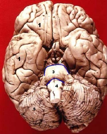
Corpora quadrigemina
The corpora quadrigemina ("quadruplet bodies") are four solid lobes on the dorsal side of the cerebral aqueduct, where the superior posterior pair are called the superior colliculi and the inferior posterior pair are called the inferior colliculi. The homologous structures are called optic lobes in some lower vertebrates (fishes, amphibians, and birds) where they integrate sensory information from the eyes and certain auditory reflexes.
The four solid lobes help to decussate several fibres of the optic nerve. However, some fibers also show ipsilateral arrangement (i.e., they run parallel on the same side without decussating.)
The superior colliculus is involved with saccadic eye movements; while the inferior is a synapsing point for sound information. The trochlear nerve comes out of the posterior surface of the midbrain, below the inferior colliculus.
Cerebral peduncle
The cerebral peduncles are paired structures, present on the ventral side of the cerebral aqueduct, and they further carry tegmentum on the dorsal side and cresta or pes on the ventral side, and both of them accommodate the corticospinal tract fibres, from the internal capsule (i.e., ascending + descending tracts = longitudinal tract.) the middle part of cerebral peduncles carry substantia nigra (literally "Black Matter"), which is a type of basal nucleus. It is the only part of the brain that carries melanin pigment.
Between the peduncles is the interpeduncular fossa, which is a cistern filled with cerebrospinal fluid. The oculomotor nerve comes out between the peduncles, and the trochlear nerve is visible wrapping around the outside of the peduncles. The oculomotor is responsible for pupil constriction (parasympathetic) and certain eye movements.
Anatomical features of cross-sections through the midbrain
The midbrain is usually sectioned at the level of the superior and inferior colliculi.
One mnemonic for remembering the structures of the midbrain involves visualizing the mesencephalic cross-section as an upside down bear face. The two red nuclei are the eyes of the bear and the cerebral crura are the ears. The tectum is the chin and the cerebral peduncles are the face and ears.
Development
During embryonic development, the midbrain arises from the second vesicle, also known as the mesencephalon, of the neural tube. Unlike the other two vesicles, the forebrain and hindbrain, the midbrain remains undivided for the remainder of neural development. It does not split into other brain areas. while the forebrain, for example, divides into the telencephalon and the diencephalon.
Throughout embryonic development, the cells within the midbrain continually multiply and compress the still-forming cerebral aqueduct. Partial or total obstruction of the cerebral aqueduct during development can lead to congenital hydrocephalus.
Function
The mesencephalon is considered part of the brainstem. Its substantia nigra is closely associated with motor system pathways of the basal ganglia. The human mesencephalon is archipallian in origin, meaning that its general architecture is shared with the most ancient of vertebrates. Dopamine produced in the substantia nigra and ventral tegmental area plays a role in excitation, motivation and habituation of species from humans to the most elementary animals such as insects. Laboratory house mice from lines that have been selectively bred for high voluntary wheel running have enlarged midbrains. The midbrain helps to relay information for vision and hearing.