
Latin diencephalon Code TH H3.11.03.5.00001 NeuroLex ID Diencephalon | MeSH A08.186.211.730.385 NeuroNames hier-271 | |
 | ||
TA A14.1.03.007A14.1.08.001 | ||
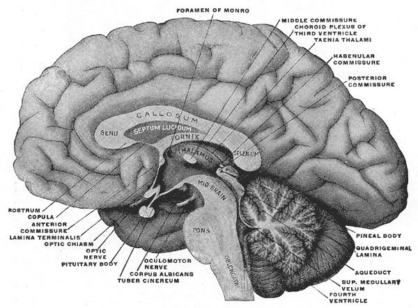
The diencephalon is part of the prosencephalon (forebrain), which develops from the foremost primary cerebral vesicle. The prosencephalon differentiates into a caudal diencephalon and rostral telencephalon. The cerebral hemispheres develop from the sides of the telencephalon, each containing a lateral ventricle. The diencephalon consists of structures that are lateral to the third ventricle, and includes the thalamus, the hypothalamus, the epithalamus and the subthalamus.
Contents
Structure
The diencephalon consists of the following structures:
Attachments
The optic nerve (CNII) attaches to the diencephalon. The optic nerve is a sensory (afferent) nerve responsible for vision; it runs from the eye through the optic canal in the skull and attaches to the diencephalon. The retina itself is derived from the optic cup, a part of the embryonic diencephalon.
Function
The diencephalon is the region of the embryonic vertebrate neural tube that gives rise to posterior forebrain structures including the thalamus, hypothalamus, posterior portion of the pituitary gland, and pineal gland. The hypothalamus performs numerous vital functions, most of which relating directly or indirectly to the regulation of visceral activities by way of other brain regions and the autonomic nervous system.