
Part of Brain stem NeuroNames hier-538 | NeuroLex ID Pons | |
 | ||
Vein transverse and lateral pontine veins MeSH A08.186.211.132.810.428.600 | ||
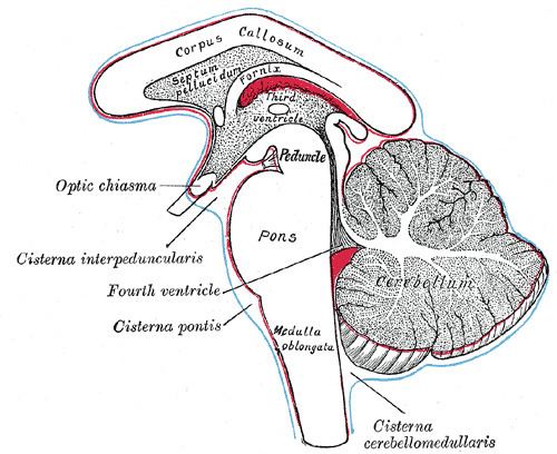
The pons is part of the brainstem, and in humans and other bipeds lies between the midbrain (above) and the medulla oblongata (below) and in front of the cerebellum.
Contents
The pons is also called the pons Varolii ("bridge of Varolius"), after the Italian anatomist and surgeon Costanzo Varolio (1543–75). This region of the brainstem includes neural pathways or tracts that conduct signals from the brain down to the cerebellum and medulla, and tracts that carry the sensory signals up into the thalamus.
The pons in humans measures about 2.5 centimetres (0.98 in) in length. Most of it appears as a broad anterior bulge rostral to the medulla. Posteriorly, it consists mainly of two pairs of thick stalks called cerebellar peduncles. They connect the cerebellum to the pons and midbrain.
The pons contains nuclei that relay signals from the forebrain to the cerebellum, along with nuclei that deal primarily with sleep, respiration, swallowing, bladder control, hearing, equilibrium, taste, eye movement, facial expressions, facial sensation, and posture.
Within the pons is the pneumotaxic center consisting of the subparabrachial and the medial parabrachial nuclei. This center regulates the change from inhalation to exhalation.
The pons is implicated in sleep paralysis, and may also play a role in generating dreams.
Structure
The pons can be broadly divided into two parts: the basilar part of the pons, located ventrally, and the pontine tegmentum, located dorsally.
Development
During embryonic development, the metencephalon develops from the rhombencephalon and gives rise to two structures: the pons and the cerebellum. The alar plate produces sensory neuroblasts, which will give rise to the solitary nucleus and its special visceral afferent (SVA) column; the cochlear and vestibular nuclei, which form the special somatic afferent (SSA) fibers of the vestibulocochlear nerve, the spinal and principal trigeminal nerve nuclei, which form the general somatic afferent column (GSA) of the trigeminal nerve, and the pontine nuclei which relays to the cerebellum.
Basal plate neuroblasts give rise to the abducens nucleus, which forms the general somatic efferent fibers (GSE); the facial and motor trigeminal nuclei, which form the special visceral efferent (SVE) column, and the superior salivatory nucleus, which forms the general visceral efferent fibers of the facial nerve.
Nucleus
A number of cranial nerve nuclei are present in the pons:
Function
The functions of these four cranial nerves (V-VIII) include sensory roles in hearing, equilibrium, and taste, and in facial sensations such as touch and pain, as well as motor roles in eye movement, facial expressions, chewing, swallowing, and the secretion of saliva and tears.
Clinical significance
Evolution
The pons first evolved as an offshoot of the medullary reticular formation. Since lampreys possess a pons, it has been argued that it must have evolved as a region distinct from the medulla by the time the first agnathans appeared, 505 million years ago.