
Latin ventriculus quartus NeuroNames hier-617 TA A14.1.05.701 | MeSH A08.186.211.276.500 NeuroLex ID Fourth ventricle FMA 78469 | |
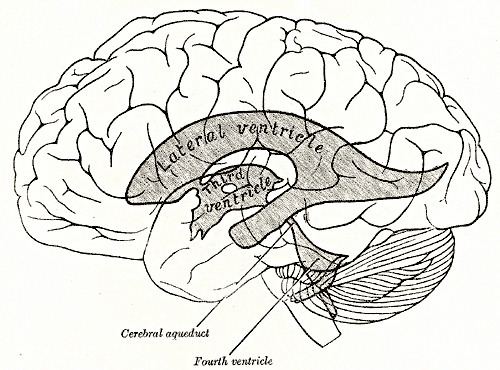 | ||
The fourth ventricle is one of the four connected fluid-filled cavities within the human brain. These cavities, known collectively as the ventricular system, consist of the left and right lateral ventricles, the third ventricle, and the fourth ventricle. The fourth ventricle extends from the cerebral aqueduct (aqueduct of Sylvius) to the obex, and is filled with cerebrospinal fluid (CSF).
Contents
The fourth ventricle has a characteristic diamond shape in cross-sections of the human brain. It is located within the pons or in the upper part of the medulla. CSF entering the fourth ventricle through the cerebral aqueduct can exit to the subarachnoid space of the spinal cord through two lateral apertures and a single, midline median aperture.
Roof and floor
The fourth ventricle has a "roof" dorsally a "floor" ventrally, and side walls formed by the cerebellar peduncles. The roof of the fourth ventricle is formed by the cerebellum (superior and inferior medullary vela). The fastigium, (Latin for summit), is used to refer to the peak of the fourth ventricle.The fastigial nucleus lies immediately above the roof of the fourth ventricle.
The floor of the fourth ventricle is formed by the rhomboid fossa. Among the prominent features of the floor of the fourth ventricle are the:
- facial colliculus: formed by the internal part of the facial nerve as it loops around the abducens nucleus in the lower pons;
- sulcus limitans: which represents the border between the alar plate and the basal plate of the developing neural tube;
- obex: represents the caudal tip of the fourth ventricle; the obex is also a marker for the level of the foramen magnum of the skull and therefore is a marker for the imaginary dividing line between the medulla and spinal cord.
- "median sulcus" - divides the floor into right and left halves. It extends from cerebral aqueduct of the midbrain to central canal of the spinal cord.
- "stria medullaris" - fibers derived from arcuate nuclei, which emerge from the median sulcus and run transversely across the floor to enter into the inferior cerebellar penducle.
- "medial eminence" - elevations on either side of the median sulcus.
- "sulcus limitans" - medial eminence is laterally bounded by sulcus limitans.
- "vestibular area" - lateral to sulcus limitans vestibular nuclei is overlied by this.
- The upper end of the sulcus limitans widens into a triangular depression called "superior fovea". Above the superior fovea sulcus limitans presents a flattened grey area called "locus ceruleus".
- The lower end of the sulcus limitans widens into a triangular depression called "Inferior fovea".
- Other features are the hypoglossal trigone and the vagal trigone.
Development
The ventricular system including the fourth ventricle, develops from the central canal of the neural tube. Specifically, the fourth ventricle originates from the portion of the tube that is present in the developing rhombencephalon. During the first trimester of pregnancy the central canal expands into the lateral, third and fourth ventricles, connected by thinner channels.Choroid plexuses appear in the ventricles which produce cerebrospinal fluid. If the flow of fluid is blocked ventricles may become enlarged and cause hydrocephalus.
Clinical significance
The fourth ventricle is a common location of an intracranial ependymomal tumour.