
Group Group I (dsDNA) | Order Herpesvirales Subfamily Alphaherpesvirinae Rank Genus | |
 | ||
Lower classifications Human herpesvirus 2, Human herpesvirus 1 | ||
Herpes simplex virus
Herpes simplex virus 1 and 2 (HSV-1 and HSV-2), also known as human herpesvirus 1 and 2 (HHV-1 and HHV-2), are two members of the herpesvirus family, Herpesviridae, that infect humans. Both HSV-1 (which produces most cold sores) and HSV-2 (which produces most genital herpes) are ubiquitous and contagious. They can be spread when an infected person is producing and shedding the virus.
Contents
- Herpes simplex virus
- Transmission
- Viral structure
- Cellular entry
- Genetic inoculation
- Immune evasion
- Replication
- Latent infection
- Evolution
- Treatment
- Alzheimers disease
- Multiplicity reactivation
- Use as an anti cancer agent
- Use in neuronal connection tracing
- Other related outcomes
- Research
- References
In simple terms, herpes simplex 1 is most commonly known as a "cold sore," while herpes simplex 2 is the one known by the public as "herpes," or "genital herpes." Herpes simplex 1 is known to infect about 95% of the human populace, and is treated less seriously than herpes simplex 2, even though both are incurable.
Symptoms of herpes simplex virus infection include watery blisters in the skin or mucous membranes of the mouth, lips, nose or genitals. Lesions heal with a scab characteristic of herpetic disease. Sometimes, the viruses cause very mild or atypical symptoms during outbreaks. However, they can also cause more troublesome forms of herpes simplex. As neurotropic and neuroinvasive viruses, HSV-1 and -2 persist in the body by becoming latent and hiding from the immune system in the cell bodies of neurons. After the initial or primary infection, some infected people experience sporadic episodes of viral reactivation or outbreaks. In an outbreak, the virus in a nerve cell becomes active and is transported via the neuron's axon to the skin, where virus replication and shedding occur and cause new sores. It is one of the most common sexually transmitted infections.
Transmission
HSV-1 and -2 are transmitted by contact with an infected area of the skin during reactivations of the virus. Herpes simplex virus (HSV)-2 is periodically shed in the human genital tract, most often asymptomatically, and most sexual transmissions occur during asymptomatic shedding. Asymptomatic reactivation means that the virus causes atypical, subtle or hard to notice symptoms that are not identified as an active herpes infection. In one study, daily genital swab samples found HSV-2 at a median of 12–28% of days among those who have had an outbreak, and 10% of days among those suffering from asymptomatic infection, with many of these episodes occurring without visible outbreak ("subclinical shedding").
In another study, 73 subjects were randomized to receive valaciclovir 1 g daily or placebo for 60 days each in a 2-way crossover design. A daily swab of the genital area was self-collected for HSV-2 detection by polymerase chain reaction, in order to compare the effect of valaciclovir 1 g once daily for 60 days versus placebo on asymptomatic viral shedding in immunocompetent, HSV-2 seropositive subjects without a history of symptomatic genital herpes infection. The study found that valaciclovir significantly reduced shedding during subclinical days compared to placebo, showing a 71% reduction. 84% of subjects had no shedding while receiving valaciclovir versus 54% of subjects on placebo. 88% of patients treated with valaciclovir had no recognized signs or symptoms versus 77% for placebo.
For HSV-2, subclinical shedding may account for most of the transmission. Studies on discordant partners (one infected with HSV-2, one not) show that the transmission rate is approximately 5 per 10,000 sexual contacts. (Effect of Condoms on Reducing the Transmission of Herpes Simplex Virus Type 2 From Men to Women. A Wald, AGM Langenberg, K Link, et al. JAMA. 2001;285(24):3197) Atypical symptoms are often attributed to other causes such as a yeast infection. HSV-1 is often acquired orally during childhood. It may also be sexually transmitted, including contact with saliva, such as kissing and mouth-to-genital contact (oral sex). HSV-2 is primarily a sexually transmitted infection, but rates of HSV-1 genital infections are increasing.
Both viruses may also be transmitted vertically during childbirth, although the real risk is very low. The risk of infection is minimal if the mother has no symptoms or exposed blisters during delivery. The risk is considerable when the mother is infected with the virus for the first time during late pregnancy.
Herpes simplex viruses can affect areas of skin exposed to contact with an infected person. An example of this is herpetic whitlow which is a herpes infection on the fingers. This was a common affliction of dental surgeons prior to the routine use of gloves when conducting treatment on patients.
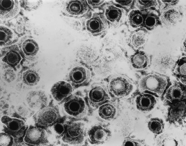
Viral structure
Animal herpes viruses all share some common properties. The structure of herpes viruses consists of a relatively large double-stranded, linear DNA genome encased within an icosahedral protein cage called the capsid, which is wrapped in a lipid bilayer called the envelope. The envelope is joined to the capsid by means of a tegument. This complete particle is known as the virion. HSV-1 and HSV-2 each contain at least 74 genes (or open reading frames, ORFs) within their genomes, although speculation over gene crowding allows as many as 84 unique protein coding genes by 94 putative ORFs. These genes encode a variety of proteins involved in forming the capsid, tegument and envelope of the virus, as well as controlling the replication and infectivity of the virus. These genes and their functions are summarized in the table below.
The genomes of HSV-1 and HSV-2 are complex and contain two unique regions called the long unique region (UL) and the short unique region (US). Of the 74 known ORFs, UL contains 56 viral genes, whereas US contains only 12. Transcription of HSV genes is catalyzed by RNA polymerase II of the infected host. Immediate early genes, which encode proteins that regulate the expression of early and late viral genes, are the first to be expressed following infection. Early gene expression follows, to allow the synthesis of enzymes involved in DNA replication and the production of certain envelope glycoproteins. Expression of late genes occurs last; this group of genes predominantly encode proteins that form the virion particle.
Five proteins from (UL) form the viral capsid; UL6, UL18, UL35, UL38 and the major capsid protein UL19.
Cellular entry
Entry of HSV into a host cell involves several glycoproteins on the surface of the enveloped virus binding to their transmembrane receptors on the cell surface. Many of these receptors are then pulled inwards by the cell, which is thought to open a ring of three gHgL heterodimers stabilizing a compact conformation of the gB glycoprotein, so that it springs out and punctures the cell membrane. The envelope covering the virus particle then fuses with the cell membrane, creating a pore through which the contents of the viral envelope enters the host cell.
The sequential stages of HSV entry are analogous to those of other viruses. At first, complementary receptors on the virus and the cell surface bring the viral and cell membranes into proximity. Interactions of these molecules then form a stable entry pore through which the viral envelope contents are introduced to the host cell. The virus can also be endocytosed after binding to the receptors, and the fusion could occur at the endosome. In electron micrographs the outer leaflets of the viral and cellular lipid bilayers have been seen merged; this hemifusion may be on the usual path to entry or it may usually be an arrested state more likely to be captured than a transient entry mechanism.
In the case of a herpes virus, initial interactions occur when two viral envelope glycoprotein called glycoprotein C (gC) and glycoprotein B (gB) bind to a cell surface particle called heparan sulfate. Next, the major receptor binding protein, glycoprotein D (gD), binds specifically to at least one of three known entry receptors. These cell receptors include herpesvirus entry mediator (HVEM), nectin-1 and 3-O sulfated heparan sulfate. The nectin receptors usually produce cell-cell adhesion, to provide a strong point of attachment for the virus to the host cell. These interactions bring the membrane surfaces into mutual proximity and allow for other glycoproteins embedded in the viral envelope to interact with other cell surface molecules. Once bound to the HVEM, gD changes its conformation and interacts with viral glycoproteins H (gH) and L (gL), which form a complex. The interaction of these membrane proteins may result in a hemifusion state. gB interaction with the gH/gL complex creates an entry pore for the viral capsid. gB interacts with glycosaminoglycans on the surface of the host cell.
Genetic inoculation
After the viral capsid enters the cellular cytoplasm, it is transported to the cell nucleus. Once attached to the nucleus at a nuclear entry pore, the capsid ejects its DNA contents via the capsid portal. The capsid portal is formed by twelve copies of portal protein, UL6, arranged as a ring; the proteins contain a leucine zipper sequence of amino acids which allow them to adhere to each other. Each icosahedral capsid contains a single portal, located in one vertex. The DNA exits the capsid in a single linear segment.
Immune evasion
HSV evades the immune system through interference with MHC class I antigen presentation on the cell surface, by blocking TAP or the transporter associated with antigen processing induced by the secretion of ICP-47 by HSV. In the host cell, TAP transports digested viral antigen epitope peptides from the cytosol to the endoplasmic reticulum, allowing these epitopes to be combined with MHC class I molecules and presented on the surface of the cell. Viral epitope presentation with MHC class I is a requirement for activation of cytotoxic T-lymphocytes (CTLs), the major effectors of the cell-mediated immune response against virally-infected cells. ICP-47 prevents initiation of a CTL-response against HSV, allowing the virus to survive for a protracted period in the host.
Replication
Following infection of a cell, a cascade of herpes virus proteins, called immediate-early, early, and late, are produced. Research using flow cytometry on another member of the herpes virus family, Kaposi's sarcoma-associated herpesvirus, indicates the possibility of an additional lytic stage, delayed-late. These stages of lytic infection, particularly late lytic, are distinct from the latency stage. In the case of HSV-1, no protein products are detected during latency, whereas they are detected during the lytic cycle.
The early proteins transcribed are used in the regulation of genetic replication of the virus. On entering the cell, an α-TIF protein joins the viral particle and aids in immediate-early transcription. The virion host shutoff protein (VHS or UL41) is very important to viral replication. This enzyme shuts off protein synthesis in the host, degrades host mRNA, helps in viral replication, and regulates gene expression of viral proteins. The viral genome immediately travels to the nucleus but the VHS protein remains in the cytoplasm.
The late proteins form the capsid and the receptors on the surface of the virus. Packaging of the viral particles — including the genome, core and the capsid - occurs in the nucleus of the cell. Here, concatemers of the viral genome are separated by cleavage and are placed into pre-formed capsids. HSV-1 undergoes a process of primary and secondary envelopment. The primary envelope is acquired by budding into the inner nuclear membrane of the cell. This then fuses with the outer nuclear membrane releasing a naked capsid into the cytoplasm. The virus acquires its final envelope by budding into cytoplasmic vesicles.
Latent infection
HSVs may persist in a quiescent but persistent form known as latent infection, notably in neural ganglia. HSV-1 tends to reside in the trigeminal ganglia, while HSV-2 tends to reside in the sacral ganglia, but these are tendencies only, not fixed behavior. During latent infection of a cell, HSVs express latency associated transcript (LAT) RNA. LAT regulates the host cell genome and interferes with natural cell death mechanisms. By maintaining the host cells, LAT expression preserves a reservoir of the virus, which allows subsequent, usually symptomatic, periodic recurrences or "outbreaks" characteristic of non-latency. Whether or not recurrences are symptomatic, viral shedding occurs to infect a new host. A protein found in neurons may bind to herpes virus DNA and regulate latency. Herpes virus DNA contains a gene for a protein called ICP4, which is an important transactivator of genes associated with lytic infection in HSV-1. Elements surrounding the gene for ICP4 bind a protein known as the human neuronal protein Neuronal Restrictive Silencing Factor (NRSF) or human Repressor Element Silencing Transcription Factor (REST). When bound to the viral DNA elements, histone deacetylation occurs atop the ICP4 gene sequence to prevent initiation of transcription from this gene, thereby preventing transcription of other viral genes involved in the lytic cycle. Another HSV protein reverses the inhibition of ICP4 protein synthesis. ICP0 dissociates NRSF from the ICP4 gene and thus prevents silencing of the viral DNA.
Evolution
The herpes simplex 1 genomes can be classified into six clades. Four of these occur in East Africa, one in East Asia and one in Europe and North America. This suggests that the virus may have originated in East Africa. The most recent common ancestor of the Eurasian strains appears to have evolved ~60,000 years ago. The East Asian HSV-1 isolates have an unusual pattern that is currently best explained by the two waves of migration responsible for the peopling of Japan.
The mutation rate has been estimated to be ~1.38×10⁻⁷ substitutions/site/year. In clinical setting, the mutations in either the thymidine kinase gene or DNA polymerase gene has caused resistance to aciclovir. However, most of the mutations occur in the thymidine kinase gene rather than the DNA polymerase gene.
Treatment
For more details on treatment of herpes simplex virus, see Herpes simplex.Herpes viruses establish lifelong infections, and the virus cannot yet be eradicated from the body. Treatment usually involves general-purpose antiviral drugs that interfere with viral replication, reduce the physical severity of outbreak-associated lesions, and lower the chance of transmission to others. Studies of vulnerable patient populations have indicated that daily use of antivirals such as aciclovir and valaciclovir can reduce reactivation rates.
Alzheimer's disease
In the presence of a certain gene variation (APOE-epsilon4 allele carriers), a possible link between HSV-1 (i.e., the virus that causes cold sores or oral herpes) and Alzheimer's disease was reported in 1979. HSV-1 appears to be particularly damaging to the nervous system and increases one’s risk of developing Alzheimer’s disease. The virus interacts with the components and receptors of lipoproteins, which may lead to the development of Alzheimer's disease. This research identifies HSVs as the pathogen most clearly linked to the establishment of Alzheimer’s. According to a study done in 1997, without the presence of the gene allele, HSV-1 does not appear to cause any neurological damage or increase the risk of Alzheimer’s. However, a more recent prospective study published in 2008 with a cohort of 591 people showed a statistically significant difference between patients with antibodies indicating recent reactivation of HSV and those without these antibodies in the incidence of Alzheimer's disease, without direct correlation to the APOE-epsilon4 allele. It should be noted that the trial had a small sample of patients who did not have the antibody at baseline, so the results should be viewed as highly uncertain. In 2011 Manchester University scientists showed that treating HSV1-infected cells with antiviral agents decreased the accumulation of β-amyloid and P-tau, and also decreased HSV-1 replication.
Multiplicity reactivation
Multiplicity reactivation (MR) is the process by which viral genomes containing inactivating damage interact within an infected cell to form a viable viral genome. MR was originally discovered with the bacterial virus bacteriophage T4, but was subsequently also found with pathogenic viruses including influenza virus, HIV-1, adenovirus simian virus 40, vaccinia virus, reovirus, poliovirus and herpes simplex virus.
When HSV particles are exposed to doses of a DNA damaging agent that would be lethal in single infections, but are then allowed to undergo multiple infection (i.e. two or more viruses per host cell), MR is observed. Enhanced survival of HSV-1 due to MR occurs upon exposure to different DNA damaging agents, including methyl methanesulfonate, trimethylpsoralen (which causes inter-strand DNA cross-links), and UV light. After treatment of genetically marked HSV with trimethylpsoralen, recombination between the marked viruses increases, suggesting that trimethylpsoralen damage stimulates recombination. MR of HSV appears to partially depend on the host cell recombinational repair machinery since skin fibroblast cells defective in a component of this machinery (i.e. cells from Bloom’s syndrome patients) are deficient in MR. These observations suggest that MR in HSV infections involves genetic recombination between damaged viral genomes resulting in production of viable progeny viruses. HSV-1, upon infecting host cells, induces inflammation and oxidative stress. Thus it appears that the HSV genome may be subjected to oxidative DNA damage during infection, and that MR may enhance viral survival and virulence under these conditions.
Use as an anti-cancer agent
Herpes simplex virus is considered as a potential therapy for cancer and has been extensively clinically tested to assess its oncolytic (cancer killing) ability. Interim overall survival data from Amgen's phase 3 trial of a genetically-attenuated herpes virus suggests efficacy against melanoma.
Use in neuronal connection tracing
Herpes simplex virus is also used as a transneuronal tracer defining connections among neurons by virtue of traversing synapses.
Other related outcomes
Herpes simplex virus is likely the most common cause of Mollaret's meningitis, and, in worse case scenarios, can lead to a potentially fatal case of herpes simplex encephalitis.
Research
There exist commonly used vaccines to some herpesviruses, but only veterinary, such as HVT/LT (Turkey herpesvirus vector laryngotracheitis vaccine). However, it prevents atherosclerosis (which histologically mirrors atherosclerosis in humans) in target animals vaccinated.