
Precursor Septum secundum Acronym(s) PFO TA A12.1.01.007 | System Cardiovascular system MeSH A07.541.459 FMA 86043 | |
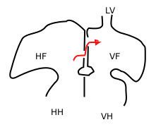 | ||
In the fetal heart, the foramen ovale (/fəˈreɪmən oʊˈvæli, -mɛn-, -ˈvɑː-, -ˈveɪ-/), also foramen Botalli, ostium secundum of Born or falx septi, allows blood to enter the left atrium from the right atrium. It is one of two fetal cardiac shunts, the other being the ductus arteriosus (which allows blood that still escapes to the right ventricle to bypass the pulmonary circulation). Another similar adaptation in the fetus is the ductus venosus. In most individuals, the foramen ovale closes at birth. It later forms the fossa ovalis.
Contents
Function
A fetus receives oxygen not from its lungs, but from the mother's oxygen-rich blood via the placenta. Oxygenated blood from the placenta travels through the umbilical cord to the right atrium of the fetal heart. As the fetal lungs are non-functional at this time, it is more efficient for the blood to bypass them. This is accomplished through 2 cardiac shunts. The first is the foramen ovale which shunts blood from the right atrium to the left atrium. The second is the ductus arteriosus which shunts blood from the pulmonary artery (which, after birth, carries blood from the right side of the heart to the lungs) to the descending aorta.
Development
The foramen ovale forms in the late fourth week of gestation. Initially the atria are separated from one another by the septum primum except for a small opening in the septum, the ostium primum. As the septum primum grows, the ostium primum narrows and eventually closes. Before it does so, bloodflow from the inferior vena cava wears down a portion of the septum primum, forming the ostium secundum. Some embryologists postulate that the ostium secundum may be formed through programmed cell death.
The ostium secundum provides communication between the atria after the ostium primum closes completely. Subsequently, a second wall of tissue, the septum secundum, grows over the ostium secundum in the right atrium. Bloodflow then only passes from the right to left atrium by way of a small passageway in the septum secundum and then through the ostium secundum. This passageway is called the foramen ovale.
Closure
Normally this opening closes at birth. When the lungs become functional at birth, the pulmonary pressure decreases and the left atrial pressure exceeds that of the right. This forces the septum primum against the septum secundum, functionally closing the foramen ovale. In time the septa eventually fuse, leaving a remnant of the foramen ovale, the fossa ovalis.
Clinical significance
In about 25% of adults the foramen ovale does not close completely, but remains as a small patent foramen ovale ("PFO"). In most of these individuals, the PFO causes no problems and remains undetected throughout life.
PFO has long been studied because of its role in paradoxical embolism (an embolism that travels from the venous side to the arterial side). This may lead to a stroke or transient ischemic attack. Transesophageal echocardiogramphy is considered the most accurate investigation to demonstrate a patent foramen ovale. A patent foramen ovale may also be an incidental finding.