
 | ||
Human fertilization is the union of a human egg and sperm, usually occurring in the ampulla of the fallopian tube. The result of this union is the production of a zygote cell, or fertilized egg, initiating prenatal development. Scientists discovered the dynamics of human fertilization in the nineteenth century.
Contents
- Corona radiata
- Cone of attraction and perivitelline membrane
- Sperm preparation
- Zona pellucida
- Cortical reaction
- Fusion
- Cell membranes
- Transformations
- Replication
- Mitosis
- Fertilization age
- Diseases
- References
The process of fertilization involves a sperm fusing with an ovum. The most common sequence begins with ejaculation during copulation, follows with ovulation, and finishes with fertilization. Various exceptions to this sequence are possible, including artificial insemination, in vitro fertilization, external ejaculation without copulation, or copulation shortly after ovulation. Upon encountering the secondary oocyte, the acrosome of the sperm produces enzymes which allow it to burrow through the outer jelly coat of the egg. The sperm plasma then fuses with the egg's plasma membrane, the sperm head disconnects from its flagellum and the egg travels down the Fallopian tube to reach the uterus.
In vitro fertilization (IVF) is a process by which egg cells are fertilized by sperm outside the womb, in vitro.
Corona radiata
The sperm binds through the corona radiata, a layer of follicle cells on the outside of the secondary oocyte. Fertilization occurs when the nucleus of both a sperm and an egg fuse to form a diploid cell, known as zygote. The successful fusion of gametes forms a new organism.
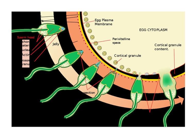
Cone of attraction and perivitelline membrane
Where the spermatozoon is about to pierce, the yolk (ooplasm) is drawn out into a conical elevation, termed the cone of attraction or reception cone. Once the spermatozoon has entered, the peripheral portion of the yolk changes into a membrane, the perivitelline membrane, which prevents the passage of additional spermatozoa.
Sperm preparation
At the beginning of the process, the sperm undergoes a series of changes, as freshly ejaculated sperm is unable or poorly able to fertilize. The sperm must undergo capacitation in the female's reproductive tract over several hours, which increases its motility and destabilizes its membrane, preparing it for the acrosome reaction, the enzymatic penetration of the egg's tough membrane, the zona pellucida, which surrounds the oocyte.
Zona pellucida
After binding to the corona radiata the sperm reaches the zona pellucida, which is an extra-cellular matrix of glycoproteins. A special complementary molecule on the surface of the sperm head binds to a ZP3 glycoprotein in the zona pellucida. This binding triggers the acrosome to burst, releasing enzymes that help the sperm get through the zona pellucida.
Some sperm cells consume their acrosome prematurely on the surface of the egg cell, facilitating the penetration by other sperm cells. As a population, sperm cells have on average 50% genome similarity so the premature acrosomal reactions aid fertilization by a member of the same cohort. It may be regarded as a mechanism of kin selection.
Recent studies have shown that the egg is not passive during this process.
Cortical reaction
Once the sperm cells find their way past the zona pellucida, the cortical reaction occurs. Cortical granules inside the secondary oocyte fuse with the plasma membrane of the cell, causing enzymes inside these granules to be expelled by exocytosis to the zona pellucida. This in turn causes the glyco-proteins in the zona pellucida to cross-link with each other — i.e. the enzymes cause the ZP2 to hydrolyse into ZP2f — making the whole matrix hard and impermeable to sperm. This prevents fertilization of an egg by more than one sperm. The cortical reaction and acrosome reaction are both essential to ensure that only one sperm will fertilize an egg.
Fusion
After the sperm enters the cytoplasm of the oocyte (also called ovocyte), the tail and the outer coating of the sperm disintegrate and the cortical reaction takes place, preventing other sperm from fertilizing the same egg. The oocyte now undergoes its second meiotic division producing the haploid ovum and releasing a polar body. The sperm nucleus then fuses with the ovum, enabling fusion of their genetic material.
Cell membranes
The cell membranes of the secondary oocyte and sperm fuse.
Transformations
In preparation for the fusion of their genetic material both the oocyte and the sperm undergo transformations as a reaction to the fusion of cell membranes.
The oocyte completes its second meiotic division. This results in a mature ovum. The nucleus of the oocyte is called a pronucleus in this process, to distinguish it from the nuclei that are the result of fertilization.
The sperm's tail and mitochondria degenerate with the formation of the male pronucleus. This is why all mitochondria in humans are of maternal origin. Still, a considerable amount of RNA from the sperm is delivered to the resulting embryo and likely influences embryo development and the phenotype of the offspring.
Replication
The pronuclei migrate toward the center of the oocyte, rapidly replicating their DNA as they do so to prepare the zygote for its first mitotic division.
Mitosis
Usually 23 chromosomes from spermatozoon and 23 chromosomes from egg cell fuse (half of spermatozoons carry X chromosome and the other half Y chromosome). Their membranes dissolve, leaving no barriers between the male and female chromosomes. During this dissolution, a mitotic spindle forms between them. The spindle captures the chromosomes before they disperse in the egg cytoplasm. Upon subsequently undergoing mitosis (which includes pulling of chromatids towards centrioles in anaphase) the cell gathers genetic material from the male and female together. Thus, the first mitosis of the union of sperm and oocyte is the actual fusion of their chromosomes. During this process also crossing-over occurs.
Each of the two daughter cells resulting from that mitosis has one replica of each chromatid that was replicated in the previous stage. Thus, they are genetically identical.
Fertilization age
Fertilization is the event most commonly used to mark the zero point in descriptions of prenatal development of the embryo or fetus. The resultant age is known as fertilization age, fertilizational age, embryonic age, fetal age or (intrauterine) developmental (IUD) age.
Gestational age, in contrast, takes the beginning of the last menstrual period (LMP) as the zero point. By convention, gestational age is calculated by adding 14 days to fertilization age and vice versa. In fact, however, fertilization usually occurs within a day of ovulation, which, in turn, occurs on average 14.6 days after the beginning of the preceding menstruation (LMP). There is also considerable variability in this interval, with a 95% prediction interval of the ovulation of 9 to 20 days after menstruation even for an average woman who has a mean LMP-to-ovulation time of 14.6. In a reference group representing all women, the 95% prediction interval of the LMP-to-ovulation is 8.2 to 20.5 days.
Fertilization age is sometimes used postnatally (after birth) as well to estimate various risk factors. For example, it is a better predictor than postnatal age for risk of intraventricular hemorrhage in premature babies treated with extracorporeal membrane oxygenation.
Diseases
Various disorders can arise from defects in the fertilization process.
However, some researchers have found that in rare pairs of fraternal twins, their origin might have been from the fertilization of one egg cell from the mother and eight sperm cells from the father. This possibility has been investigated by computer simulations of the fertilization process.