
Order Enterobacteriales Rank Species | Phylum Proteobacteria Scientific name Enterobacter cloacae Higher classification Enterobacter | |
 | ||
Similar Enterobacter, Bacteria, Citrobacter freundii, Enterobacter aerogenes, Citrobacter | ||
Enterobacter cloacae
Enterobacter cloacae is a clinically significant Gram-negative, facultatively-anaerobic, rod-shaped bacterium.
Contents
- Enterobacter cloacae
- Microbiology
- Industrial use
- Safety
- Genomics
- Clinical significance
- Species of the E cloacae complex
- References
Microbiology
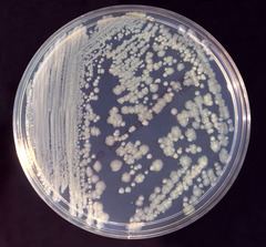
In microbiology labs, E. cloacae is frequently grown at 30 °C on nutrient agar or broth or at 35 °C in tryptic soy broth. It is a rod-shaped, Gram-negative bacterium, is facultatively anaerobic, and bears peritrichous flagella. It is oxidase-negative and catalase-positive.
Industrial use
Enterobacter cloacae has been used in a bioreactor-based method for the biodegradation of explosives and in the biological control of plant diseases.
Safety
E. cloacae is considered a biosafety level 1 organism in the United States and level 2 in Canada.
Genomics
A draft genome sequence of Enterobacter cloacae subsp. cloacae was announced in 2012. The bacteria used in the study were isolated from giant panda feces.
Clinical significance
Enterobacter cloacae is a member of the normal gut flora of many humans and is not usually a primary pathogen. It is sometimes associated with urinary tract and respiratory tract infections. Treatment with cefepime and gentamicin has been reported.
A 2012 study in which Enterobacter cloacae transplanted into previously germ-free mice resulted in increased obesity when compared with germ-free mice fed an identical diet, suggesting a link between obesity and the presence of Enterobacter gut flora.
Species of the E. cloacae complex
E. cloacae was described for the first time in 1890 by Jordan[201] as Bacillus cloacae, and then underwent numerous taxonomical changes, becoming 'Bacterium cloacae' in 1896 (Lehmann and Neumann), Cloaca cloacae in 1919 (Castellani and Chalmers), it was identified as 'Aerobacter cloacae' in 1923 (Bergey et al.), Aerobacter cloacae in 1958 (Hormaeche and Edwards) and E. cloacae in 1960 (Hormaeche and Edwards), by which it is still known today.[7] E. cloacae is ubiquitous in terrestrial and aquatic environments (water, sewage, soil and food). These strains occur as commensal microflora in the intestinal tracts of humans and animals[1] and play an important role as pathogens in plants and insects. This diversity of habitats is mirrored by the genetic variety of the nomenspecies E. cloacae.[6] E. cloacae is also an important nosocomial pathogen responsible for bacteremia and lower respiratory tract, urinary tract and intra-abdominal infections, as well as endocarditis, septic arthritis, osteomyelitis and skin and soft tissue infections. The skin and the GI tract are the most common sites through which E. cloacae can be contracted.[1,29]
E. cloacae tends to contaminate various medical, intravenous and other hospital devices. Nosocomial outbreaks have also been associated with colonization of certain surgical equipment and operative cleaning solutions. Another potential reservoir for nosocomial bacteremia is the heparin solution used to irrigate certain intravascular devices continually. This fluid had been implicated as a reservoir for outbreaks of device-associated bacteremia in several instances.[30]
In recent years, E. cloacae has emerged as one of the most commonly found nosocomial pathogen in neonatal units, with several outbreaks of infection being reported.[31] In 1998, van Nierop et al. reported an outbreak in a neonatal intensive care unit with nine deaths,[32] and in 2003, Kuboyama et al. reported three outbreaks with 42 systemic infections and a mortality of 34%.[33] This microorganism may be transmitted to neonates through contaminated intravenous fluids, total parenteral nutrition solutions and medical equipment. Many single-clone outbreaks, probably caused by cross-transmission via healthcare workers, have been described, suggesting that inpatients can also act as a reservoir.[31] The type strains of the species are E. cloacae ATCC 49162 and 13047. This latter strain is the first complete genome sequence of the E. cloacae species and the type strain is E. cloacae subsp. cloacae.
The complete E. cloacae subsp. cloacae ATCC 13047 genome contains a single circular chromosome of 5,314,588 bp and two circular plasmids, pECL_A and pECL_B, of 200,370 and 85,650 bp (GenBank accession numbers CP001918, CP001919 and CP001920, respectively).[34]
The other genomes of E. cloacae that have been sequenced are deposited in GenBank under accession numbers CP002272, CP002886, FP929040 and AGSY00000000.
E. asburiae is named after Mary Alyce Fife-Asbury, an American bacteriologist who made many important contributions to the classification of Enterobacteriaceae, particularly in describing new Klebsiella and Salmonella serotypes,[35–37] new genera and new species.[38–42] E. asburiae sp. nov. was described in 1986 based on the enteric group 17.[43] This group was defined in 1978 as a group of biochemically similar strains isolated from different human specimens[44] and sent to the CDC. Before the designation of 'enteric group 17', these strains had been reported as unidentified or atypical Citrobacter or Enterobacter strains.[44] After several studies, it was shown that these strains represent a single new species in the genus Enterobacter, which was named E. asburiae.
E. asburiae strains have been isolated from the soil and implicated in the mobilization of phosphate for plant nutrition from calcium phosphate, but most E. asburiae species have been isolated from human sources. The type strain of the species E. asburiae is ATCC 35953 and was isolated from lochia exudates of a 22-year-old woman in the USA.[43] The only sequenced strain of E. asburiae is LF7a, which contains a circular DNA (4,812,833 bp) and two circular plasmids, pENTAS01 (166,725 bp) and pENTAS02 (32,574 bp), which were submitted by Lucas et al. in 2011 to the US DOE Joint Genome Institute (CA, USA; GenBank accession numbers CP003026.1, CP003027.1 and CP003028.1, respectively).
E. hormaechei is named after Estenio Hormaeche, a Uruguayan microbiologist who (with PR Edwards) proposed and defined the genus Enterobacter.[7] The name E. hormaechei was formerly called enteric group 75, which contained 11 strains that were sent to the CDC for identification between 1973 and 1984. Twelve additional strains were received from 1985 to 1987, three of which were blood isolates. E. hormaechei was first described on the basis of 23 isolates sent to the CDC for identification. At that time, they could not be assigned to a species since they were negative in the D-sorbitol and melibiose tests and did not fit the biochemical profile of any established Enterobacter species. The species E. hormaechei was proposed to be lactose-, D-sorbitol-, raffinose-, melibiose- and esculin-negative and 87% dulcitol-positive. These species were originally defined by O'Hara et al. when a large hybridization group of enteric organisms was isolated and found to be associated with bloodstream infections.[10]
The type strain of E. hormaechei is ATCC 49162 and was isolated from the sputum of a man in California in 1977.[10] The whole-genome shotgun sequencing project was submitted in 2011 to the Human Genome Sequencing Center (TX, USA; GenBank accession number AFHR00000000).
E. hormaechei consists of three different subspecies: E. hormaechei subsp. oharae, E. hormaechei subsp. hormaechei and E. hormaechei subsp. steigerwaltii, which corresponds to genetic clusters VI, VII and VIII, respectively.[8] The differentiation of these subspecies is based on their particular properties and biochemical tests.[11]
E. hormaechei is commonly isolated as a nosocomial pathogen of clinical significance;[45,46] it has been reported in several outbreaks of sepsis in neonatal intensive care units in the USA[47] and in Brazil, where the outbreak originated from contaminated parenteral nutrition.[48]
E. kobei is named after Kobe City (Japan), where the type strain of this species was isolated. E. kobei was first described by Kosako et al. based on a collection of 23 strains with the general traits of E. cloacae and the common phenotypic difference of being Voges–Proskauer-negative.[49] The name E. kobei is proposed for a group of organisms referred to as NIH group 21 at the NIH, Tokyo. It was later found that NIH group 21 also resembled the CDC enteric group 69,[50] and E. kobei was compared with the latter. On the basis of DNA relatedness, both organisms could be included in a single taxon. However, the CDC enteric group 69 was described as positive in Voges–Proskauer and yellow pigmentation,[50] whereas all strains of E. kobei were Voges–Proskauer- and pigmentation-negative. These findings suggest that the relationship of both organisms is at the subspecies or biogroup level. The type strain of E. kobei is NIH 1485–1479 and was isolated by blood culture of a diabetic patient.
E. ludwigii, named after Wolfgang Ludwig, a microbiologist working in bacterial systematics[51] and who developed the ARB databases as well as making them public.[52] This description is based on the phylogenetic analyses of partial hsp60 sequence data collected in a population genetic study,[6] as well as on DNA–DNA hybridization assays and phenotypic characterizations.
The type strain EN-119T was isolated from midstream urine of an 18-year-old male patient with a nosocomial urinary tract infection while he was hospitalized at the Grosshadern University-Hospital Munich, Germany. The GenBank accession number of the 16S rDNA of strain EN-119T is AJ853891.[12]
E. nimipressuralis The species E. nimipressuralis was originally defined by Brenner et al. and was formerly called Erwinia nimipressuralis, which was isolated from nonclinical sources (e.g., elm trees with a disease called wet wood).[43] Erwinia nimipressuralis was inserted in the Approved Lists of Bacterial Names in 1980. This microorganism is biochemically similar to E. cloacae, but it is different for acid production from sucrose and raffinose, whereas E. cloacae is positive in these tests. The type strain of E. nimipressuralis is ATCC 9912 and isolated from the elm Ulmus spp. in the USA (GenBank accession number AJ567900).
E. cloacae subsp.cloacae strain PR-4 was isolated and identified by 16S rDNA gene sequence with phylogenetic tree view from explosive laden soil by P Ravikumar (GenBank accession number KP261383).
E. cloacae SG208 identified as a predominant microorganism in mixed culture isolated from petrochemical sludge, IOCL, Guwahati is responsible for degradation of benzene was reported by Padhi and Gokhale (2016)[137].