
Symbol Catalase InterPro IPR011614 SCOP 7cat | Pfam PF00199 PROSITE PDOC00395 SUPERFAMILY 7cat | |
 | ||
Catalase is a common enzyme found in nearly all living organisms exposed to oxygen (such as bacteria, plants, and animals). It catalyzes the decomposition of hydrogen peroxide to water and oxygen. It is a very important enzyme in protecting the cell from oxidative damage by reactive oxygen species (ROS). Likewise, catalase has one of the highest turnover numbers of all enzymes; one catalase molecule can convert millions of hydrogen peroxide molecules to water and oxygen each second.
Contents
- Structure
- History
- Action
- Molecular mechanism
- Cellular role
- Distribution among organisms
- Clinical significance and application
- Catalase test
- Gray hair
- Interactions
- References
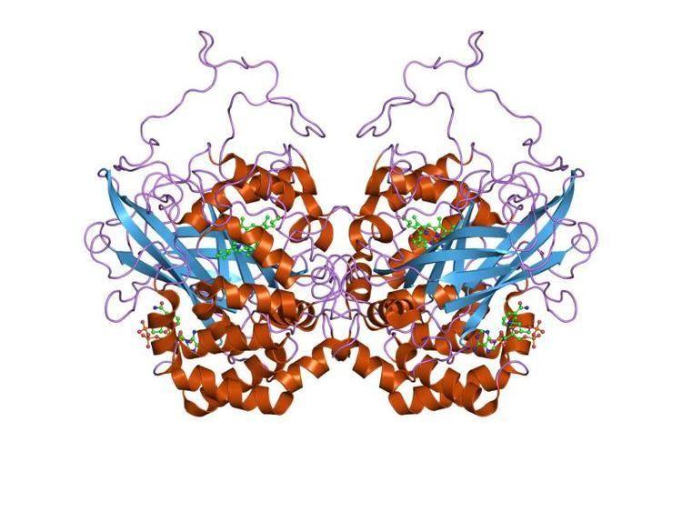
Catalase is a tetramer of four polypeptide chains, each over 500 amino acids long. It contains four porphyrin heme (iron) groups that allow the enzyme to react with the hydrogen peroxide. The optimum pH for human catalase is approximately 7, and has a fairly broad maximum (the rate of reaction does not change appreciably at pHs between 6.8 and 7.5). The pH optimum for other catalases varies between 4 and 11 depending on the species. The optimum temperature also varies by species.
Structure
Human catalase forms a tetramer composed of four subunits, each of which can be conceptually divided into four domains. The extensive hydrophobic core of each subunit is generated by an eight-stranded antiparallel b-barrel (b1-8), with nearest neighbor connectivity capped by b-barrel loops on one side and a9 on the other. A helical domain at one face of the b-barrel is composed of four C-terminal helices (a16, a17, a18, and a19) and four helices derived from residues between b4 and b5 (a4, a5, a6, and a7).
History
Catalase was not noticed until 1818 when Louis Jacques Thénard, who discovered H2O2 (hydrogen peroxide), suggested its breakdown is caused by an unknown substance. In 1900, Oscar Loew was the first to give it the name catalase, and found it in many plants and animals. In 1937 catalase from beef liver was crystallised by James B. Sumner and Alexander Dounce and the molecular weight was found in 1938.
In 1969, the amino acid sequence of bovine catalase was discovered. Then in 1981, the three-dimensional structure of the protein was revealed.
Action
The reaction of catalase in the decomposition of hydrogen peroxide in living tissue:
2 H2O2 → 2 H2O + O2The presence of catalase in a microbial or tissue sample can be tested by adding a volume of hydrogen peroxide and observing the reaction. The formation of bubbles, oxygen, indicates a positive result. This easy assay, which can be seen with the naked eye, without the aid of instruments, is possible because catalase has a very high specific activity, which produces a detectable response. Alternative splicing may result in different protein variants.
Molecular mechanism
While the complete mechanism of catalase is not currently known, the reaction is believed to occur in two stages:
H2O2 + Fe(III)-E → H2O + O=Fe(IV)-E(.+)H2O2 + O=Fe(IV)-E(.+) → H2O + Fe(III)-E + O2Here Fe()-E represents the iron center of the heme group attached to the enzyme. Fe(IV)-E(.+) is a mesomeric form of Fe(V)-E, meaning the iron is not completely oxidized to +V, but receives some "supporting electrons" from the heme ligand. This heme has to be drawn then as a radical cation (.+).As hydrogen peroxide enters the active site, it interacts with the amino acids Asn147 (asparagine at position 147) and His74, causing a proton (hydrogen ion) to transfer between the oxygen atoms. The free oxygen atom coordinates, freeing the newly formed water molecule and Fe(IV)=O. Fe(IV)=O reacts with a second hydrogen peroxide molecule to reform Fe(III)-E and produce water and oxygen. The reactivity of the iron center may be improved by the presence of the phenolate ligand of Tyr357 in the fifth iron ligand, which can assist in the oxidation of the Fe(III) to Fe(IV). The efficiency of the reaction may also be improved by the interactions of His74 and Asn147 with reaction intermediates. In general, the rate of the reaction can be determined by the Michaelis-Menten equation.
Catalase can also catalyze the oxidation, by hydrogen peroxide, of various metabolites and toxins, including formaldehyde, formic acid, phenols, acetaldehyde and alcohols. It does so according to the following reaction:
H2O2 + H2R → 2H2O + RThe exact mechanism of this reaction is not known.
Any heavy metal ion (such as copper cations in copper(II) sulfate) can act as a noncompetitive inhibitor of catalase. Furthermore, the poison cyanide is a competitive inhibitor of catalase at high concentrations of hydrogen peroxide. Arsenate acts as an activator. Three-dimensional protein structures of the peroxidated catalase intermediates are available at the Protein Data Bank. This enzyme is commonly used in laboratories as a tool for learning the effect of enzymes upon reaction rates.
Cellular role
Hydrogen peroxide is a harmful byproduct of many normal metabolic processes; to prevent damage to cells and tissues, it must be quickly converted into other, less dangerous substances. To this end, catalase is frequently used by cells to rapidly catalyze the decomposition of hydrogen peroxide into less-reactive gaseous oxygen and water molecules.
The true biological significance of catalase is not always straightforward to assess: Mice genetically engineered to lack catalase were thought to be phenotypically normal., however, A catalase deficiency may increase the likelihood of developing Obesity, fatty liver, and type 2 diabetes. Some humans have very low levels of catalase (acatalasia), yet show few ill effects. The predominant scavengers of H2O2 in normal mammalian cells are likely peroxiredoxins rather than catalase.
Human catalase works at an optimum temperature of 45 °C. In contrast, catalase isolated from the hyperthermophile archaeon Pyrobaculum calidifontis has a temperature optimum of 90 °C.
Catalase is usually located in a cellular, bipolar environment organelle called the peroxisome. Peroxisomes in plant cells are involved in photorespiration (the use of oxygen and production of carbon dioxide) and symbiotic nitrogen fixation (the breaking apart of diatomic nitrogen (N2) to reactive nitrogen atoms). Hydrogen peroxide is used as a potent antimicrobial agent when cells are infected with a pathogen. Catalase-positive pathogens, such as Mycobacterium tuberculosis, Legionella pneumophila, and Campylobacter jejuni, make catalase to deactivate the peroxide radicals, thus allowing them to survive unharmed within the host.
Catalase contributes to ethanol metabolism in the body after ingestion of alcohol, but it only breaks down a small fraction of the alcohol in the body.
Distribution among organisms
The large majority of known organisms use catalase in every organ, with particularly high concentrations occurring in the liver. One unique use of catalase occurs in the bombardier beetle. This beetle has two sets of chemicals ordinarily stored separately in its paired glands. The larger of the pair, the storage chamber or reservoir, contains hydroquinones and hydrogen peroxide, whereas the smaller of the pair, the reaction chamber, contains catalases and peroxidases. To activate the noxious spray, the beetle mixes the contents of the two compartments, causing oxygen to be liberated from hydrogen peroxide. The oxygen oxidizes the hydroquinones and also acts as the propellant. The oxidation reaction is very exothermic (ΔH = −202.8 kJ/mol) which rapidly heats the mixture to the boiling point.
Catalase is also universal among plants, and many fungi are also high producers of the enzyme.
Almost all aerobic microorganisms use catalase. It is also present in some anaerobic microorganisms, such as Methanosarcina barkeri.
According to a March 2015 Scientific American Special Report on Aging article, laboratory mice at a University of Washington laboratory which produced more catalase, which is an antioxidant, lived longer. Research on the topic both supports and cautions against the benefit of antioxidants for health effects on aging.
Clinical significance and application
Catalase is used in the food industry for removing hydrogen peroxide from milk prior to cheese production. Another use is in food wrappers where it prevents food from oxidizing. Catalase is also used in the textile industry, removing hydrogen peroxide from fabrics to make sure the material is peroxide-free.
A minor use is in contact lens hygiene - a few lens-cleaning products disinfect the lens using a hydrogen peroxide solution; a solution containing catalase is then used to decompose the hydrogen peroxide before the lens is used again. Recently, catalase has also begun to be used in the aesthetics industry. Several mask treatments combine the enzyme with hydrogen peroxide on the face with the intent of increasing cellular oxygenation in the upper layers of the epidermis.
Catalase test
The catalase test is also one of the main three tests used by microbiologists to identify species of bacteria. The presence of catalase enzyme in the test isolate is detected using hydrogen peroxide. If the bacteria possess catalase (i.e., are catalase-positive), when a small amount of bacterial isolate is added to hydrogen peroxide, bubbles of oxygen are observed.
The catalase test is done by placing a drop of hydrogen peroxide on a microscope slide. Using an applicator stick, a scientist touches the colony, and then smears a sample into the hydrogen peroxide drop.
While the catalase test alone cannot identify a particular organism, combined with other tests, such as antibiotic resistance, it can aid identification. The presence of catalase in bacterial cells depends on both the growth condition and the medium used to grow the cells.
Capillary tubes may also be used. A small amount of bacteria is collected on the end of the capillary tube (it is essential to ensure that the end is not blocked, otherwise it may present a false negative). The opposite end is then dipped into hydrogen peroxide which will draw up the liquid (through capillary action), and turned upside down, so the bacterial end is closest to the bench. A few taps of the arm should then move the hydrogen peroxide closer to the bacteria. When the hydrogen peroxide and bacteria are touching, bubbles may begin to rise, giving a positive catalase result.
Gray hair
According to recent scientific studies, low levels of catalase may play a role in the graying process of human hair. Hydrogen peroxide is naturally produced by the body and catalase breaks it down. If catalase levels decline, hydrogen peroxide cannot be broken down as well. This allows the hydrogen peroxide to bleach the hair from the inside out. This finding is under investigation in hopes of developing cosmetic treatments for graying hair based on catalase supplements.
Interactions
Catalase has been shown to interact with the ABL2 and Abl genes. Infection with the murine leukemia virus causes catalase activity to decline in the lungs, heart and kidneys of mice. Conversely, dietary fish oil increased catalase activity in the heart, and kidneys of mice.