
ICD-9-CM 282.44 DiseasesDB 3087 1373 | ICD-10 D56.1 OMIM 141900 eMedicine article/199534 | |
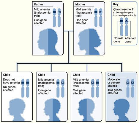 | ||
Beta thalassemias (β thalassemias) are a group of inherited blood disorders. They are forms of thalassemia caused by reduced or absent synthesis of the beta chains of hemoglobin that result in variable outcomes ranging from severe anemia to clinically asymptomatic individuals. Global annual incidence is estimated at 1 in 100,000. Beta thalassemias are caused by mutations in the HBB gene on chromosome 11, inherited in an autosomal recessive fashion. The severity of the disease depends on the nature of the mutation.
Contents
- Signs and symptoms
- Mutations
- mRNA assembly
- Diagnosis
- DNA analysis
- Beta thalassemia major
- Beta thalassemia intermedia
- Beta thalassemia minor
- Epidemiology
- Evolutionary adaptation
- References
HBB blockage over time leads to decreased beta-chain synthesis. The body's inability to construct new beta-chains leads to the underproduction of HbA. Reductions in HbA available overall to fill the red blood cells in turn leads to microcytic anemia. Microcytic anemia ultimately develops in respect to inadequate HBB for sufficient red blood cell functioning. Due to this factor, the patient may require blood transfusions to make up for the blockage in the beta-chains. Repeated blood transfusions can lead to build-up of iron overload, ultimately resulting in iron toxicity. This iron toxicity can cause various problems, including myocardial siderosis and heart failure leading to the patient’s death.
Signs and symptoms
Three main forms have been described: thalassemia major, thalassemia intermedia and thalassemia minor. All people with thalassemia are susceptible to health complications that involve the spleen (which is often enlarged and frequently removed) and gallstones. These complications are mostly found in thalassemia major and intermedia patients. Individuals with beta thalassemia major usually present within the first two years of life with severe anemia, poor growth and skeletal abnormalities during infancy. Untreated thalassemia major eventually leads to death, usually by heart failure; therefore, birth screening is very important.
Excess iron causes serious complications within the liver, heart and endocrine glands. Severe symptoms include liver cirrhosis, liver fibrosis and in extreme cases, liver cancer. Heart failure, growth impairment, diabetes and osteoporosis are life-threatening contributors brought upon by TM. The main cardiac abnormalities seen to have resulted from thalassemia and iron overload include left ventricular systolic and diastolic dysfunction, pulmonary hypertension, valveulopathies, arrhythmias and pericarditis. Increased gastrointestinal iron absorption is seen in all grades of beta thalassemia and increased red blood cell destruction by the spleen due to ineffective erythropoiesis further releases additional iron into the bloodstream.
Mutations
Two major groups of mutations can be distinguished:
mRNA assembly
Beta thalassemia is a hereditary disease affecting hemoglobin. As with about half of all hereditary diseases, an inherited mutation damages the assembly of the messenger-type RNA (mRNA) that is transcribed from a chromosome. DNA contains both the instructions (genes) for stringing amino acids together into proteins, as well as stretches of DNA that play important roles in regulating produced protein levels.
In thalassemia, an additional, contiguous length or a discontinuous fragment of non-coding instructions are included in the mRNA. This happens because the mutation obliterates the boundary between the intronic and exonic portions. Because all the coding sections may still be present, normal hemoglobin may be produced and the added material, if it produces pathology, instead disrupts regulatory functions enough to produce anemia.Hemoblogin's normal alpha and beta subunits each have an iron-containing central portion (heme) that allows the protein chain of a subunit to fold around it. Normal adult hemoglobin contains 2 alpha and 2 beta subunits. Thalassemias typically affect only the mRNAs for production of the beta chains (hence the name). Since the mutation may be a change in only a single base (a "Single Nucleotide Polymorphism"), on-going efforts seek gene therapies to make that single correction.
Diagnosis
Abdominal pain due to hypersplenism and splenic infarction and right-upper quadrant pain caused by gallstones are major clinical manifestations. However, diagnosing thalassemiæ from symptoms alone is inadequate. Physicians note these signs as associative due to this disease's complexity. The following associative signs can attest to the severity of the phenotype: pallor, poor growth, inadequate food intake, splenomegaly, jaundice, maxillary hyperplasia, dental malocclusion, cholelithiasis, systolic ejection murmur in the presence of severe anemia and pathologic fractures. Based on symptoms, tests are ordered for a differential diagnosis. These tests include complete blood count; hemoglobin electrophoresis; serum transferrin, ferritin, total iron-binding capacity; urine urobilin and urobilogen; peripheral blood smear, which may show codocytes, or target cells; hematocrit; and serum bilirubin.
DNA analysis
All beta thalassemias may exhibit abnormal red blood cells, a family history is followed by DNA analysis. This test is used to investigate deletions and mutations in the alpha- and beta-globin-producing genes. Family studies can be done to evaluate carrier status and the types of mutations present in other family members. DNA testing is not routine, but can help diagnose thalassemia and determine carrier status. In most cases the treating physician uses a clinical prediagnosis assessing anemia symptoms: fatigue, breathlessness and poor exercise tolerance. Further genetic analysis may include HPLC should routine electrophoresis prove difficult.
Beta thalassemia major
Affected children require regular lifelong blood transfusion and can have complications, which may involve the spleen. Bone marrow transplants can be curative for some children. Patients receive frequent blood transfusions that lead to or potentiate iron overload. Iron chelation treatment is necessary to prevent damage to internal organs. Advances in iron chelation treatments allow patients with thalassemia major to live long lives with access to proper treatment. Popular chelators include deferoxamine and deferiprone.
The most common patient deferoxamine complaint is that they are painful and inconvenient. The oral chelator deferasirox was approved for use in 2005 in some countries, it offers some hope with compliance at a higher cost. Bone marrow transplantation is the only cure and is indicated for patients with severe thalassemia major. Transplantation can eliminate a patient's dependence on transfusions. Absent a matching donor, a savior sibling can be conceived by preimplantation genetic diagnosis (PGD) to be free of the disease as well as to match the recipient's human leukocyte antigen (HLA) type.
Scientists at Weill Cornell Medical College have developed a gene therapy strategy that could feasibly treat both beta-thalassemia and sickle cell disease. The technology is based on delivery of a lentiviral vector carrying both the human β-globin gene and an ankyrin insulator to improve gene transcription and translation, and boost levels of β-globin production.
Beta thalassemia intermedia
Patients may require episodic blood transfusions. Transfusion-dependent patients develop iron overload and require chelation therapy to remove the excess iron. Transmission is autosomal recessive; however, dominant mutations and compound heterozygotes have been reported. Genetic counseling is recommended and prenatal diagnosis may be offered. Alleles without a mutation that reduces function are characterized as (β). Mutations are characterized as (βo) if they prevent any formation of β chains, mutations are characterized as (β+) if they allow some β chain formation to occur.
Beta thalassemia minor
Patients are often monitored without treatment. While many of those with minor status do not require transfusion therapy, they still risk iron overload, particularly in the liver. A serum ferritin test checks iron levels and can point to further treatment. Although not life-threatening on its own, it can affect quality of life due to the anemia. Minor often coexists with other conditions such as asthma and can cause iron overload of the liver and in those with non-alcoholic fatty liver disease, lead to more severe outcomes.
Epidemiology
The beta form of thalassemia is particularly prevalent among the Mediterranean peoples and this geographical association is responsible for its naming: thalassa (θάλασσα) is the Greek word for sea and haema (αἷμα) is the Greek word for blood. In Europe, the highest concentrations of the disease are found in Greece and the Turkish coastal regions. The major Mediterranean islands (except the Balearics) such as Sicily, Sardinia, Corsica, Cyprus, Malta and Crete are heavily affected in particular. Other Mediterranean peoples, as well as those in the vicinity of the Mediterranean, also have high incidence rates, including people from West Asia and North Africa. The data indicate that 15% of the Greek and Turkish Cypriots are carriers of beta-thalassaemia genes, while 10% of the population carry alpha-thalassaemia genes.
Evolutionary adaptation
The thalassemia trait may confer a degree of protection against malaria, which is or was prevalent in the regions where the trait is common, thus conferring a selective survival advantage on carriers (known as heterozygous advantage), thus perpetuating the mutation. In that respect, the various thalassemias resemble another genetic disorder affecting hemoglobin, sickle-cell disease.