
MeSH D006400 | MedlinePlus 003646 | |
 | ||
The hematocrit (Template:TIPAc-en) (Ht or HCT), also known by several other names, is the volume percentage (vol%) of red blood cells in blood. It is normally 45% for men and 40% for women. It is considered an integral part of a person's complete blood count results, along with hemoglobin concentration, white blood cell count, and platelet count. Because the purpose of red blood cells is to transfer oxygen from the lungs to body tissues, a blood sample's hematocrit—the red blood cell volume percentage—can become a point of reference of its capability of delivering oxygen. Additionally, the measure of a subject's blood sample's hematocrit levels may expose possible diseases in the subject. Anemia refers to an abnormally low hematocrit, as opposed to polycythemia, which refers to an abnormally high hematocrit. For a condition such as anemia that goes unnoticed, one way it can be diagnosed is by measuring the hematocrit levels in the blood. Both are potentially life-threatening disorders.
Contents
Names
Hematocrit is also sometimes called packed cell volume (PCV), volume of packed red cells (VPRC), or erythrocyte volume fraction (EVF). The term hematocrit comes from the Ancient Greek words haima (αἷμα, "blood") and kritēs (κριτής, "judge"). Together, hematocrit means "to separate blood". It was coined by Magnus Blix at Uppsala in 1891 as haematokrit, modeled after lactokrit, which was used in dairy farming.
Measurement methods
With modern lab equipment, the hematocrit is calculated by an automated analyzer and not directly measured. It is determined by multiplying the red cell count by the mean cell volume. The hematocrit is slightly more accurate as the PCV includes small amounts of blood plasma trapped between the red cells. An estimated hematocrit as a percentage may be derived by tripling the hemoglobin concentration in g/dL and dropping the units.
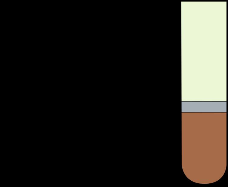
The packed cell volume (PCV) can be determined by centrifuging heparinized blood in a capillary tube (also known as a microhematocrit tube) at 10,000 RPM for five minutes. This separates the blood into layers. The volume of packed red blood cells divided by the total volume of the blood sample gives the PCV. Since a tube is used, this can be calculated by measuring the lengths of the layers.
Another way of measuring hematocrit levels has been through optical methods such as spectrophotometry has also been developed. Through differential spectrophotometry, the differences in optical densities of a blood sample flowing through small-bore glass tubes at isosbestic wavelengths for deoxyhemoglobin and oxyhemoglobin and the product of the luminal diameter and hematocrit create a linear relationship that is used to measure hematocrit levels.
There are some risks and side effects that accompany the tests of hematocrit because blood is being extracted from subjects. Subjects may experience a more than normal amount of hemorrhaging, hematoma, fainting, and possibly infection.
While known hematocrit levels are used in detecting conditions, it may fail at times due to hematocrit being the measure of concentration of red blood cells through volume in a blood sample. It does not account for the mass of the red blood cells, and thus the changes in mass can alter a hematocrit level or go undetected while affecting a subject's condition. Additionally, there have been cases in which the blood for testing was inadvertently drawn proximal to an intravenous line that was infusing packed red cells or fluids. In these situations, the hemoglobin level in the blood sample will not be the true level for the patient because the sample will contain a large amount of the infused material rather than what is diluted into the circulating whole blood. That is, if packed red cells are being supplied, the sample will contain a large amount of those cells and the hematocrit will be artificially very high. On the converse, if saline or other fluids are being supplied, the blood sample would be diluted and the hematocrit will be artificially low.
Levels
Hematocrit, the volume percentage of red blood cells, can vary from the determining factors of the number of red blood cells. These factors can be from the age and sex of the subject. Typically, a higher hematocrit level signifies the blood sample's ability to transport oxygen, which has led to reports that an "optimal hematocrit level" possibly exists. Optimal hematocrit levels have been studied through combinations of assays on blood sample's hematocrit itself, viscosity, and hemoglobin level.
Hematocrit levels also serve as an indicator of health conditions. Thus, tests on hematocrit levels are often carried out in the process of diagnosis of such conditions and may be conducted prior to surgery. Additionally, the health conditions associated with certain hematocrit levels are the same as ones associated with certain hemoglobin levels.
As blood flows from the arterioles into the capillaries a change in pressure occurs. In order to maintain pressure, the capillaries branch off to a web of vessels that carry blood into the venules. Through this process blood undergoes micro-circulation. In micro-circulation, the Fahraeus effect will take place, resulting in a large change in hematocrit. As blood flows through the arterioles, red cells will act a feed hematocrit (Hf), while in the capillaries a tube hematocrit (Ht) occurs. In tube hematocrit plasma fills most of the vessel while the red cells travel through in somewhat of a single file line. From this stage, blood will enter the venules increasing in hematocrit, in other words the discharge hematocrit (Hd).
In large vessels with low hematocrit, viscosity dramatically drops and red cells take in a lot of energy. While in smaller vessels at the micro-circulation scale, viscosity is very high. With the increase in shear stress at the wall, a lot of energy is used to move cells.
Shear rate relations
Relationships between hematocrit, viscosity, and shear rate are important factors to put into consideration. Since blood is non-Newtonian, the viscosity of the blood is in relation to the hematocrit, and as a function of shear rate. This is important when it comes to determining shear force, since a lower hematocrit level indicates that there is a need for more force to push the red blood cells through the system. This is because shear rate is defined as the rate to which adjacent layers of fluid move in respect to each other. Plasma is a more viscous material than typically red blood cells, since they are able to adjust their size to the radius of a tube; the shear rate is purely dependent on the amount of red blood cells being forced in a vessel.
Elevated
Generally at both sea levels and high altitudes, hematocrit levels rise as children mature. These health-related causes and impacts of elevated hematocrit levels have been reported:
Hematocrit levels were also reported to be influenced by social factors that influence subjects. In the 1966–80 Health Examination Survey, there was a small rise in mean hematocrit levels in female and male adolescents that reflected a rise in annual family income. Additionally, a higher education in a parent has been put into account for a rise in mean hematocrit levels of the child.
Lowered
Lowered hematocrit levels also pose health impacts. These causes and impacts have been reported: