
Scientific name Toxoplasma gondii Higher classification Toxoplasma | Subfamily Toxoplasmatinae Domain Eukaryota | |
 | ||
Similar Apicomplexa, Cat, Neospora, Trypanosoma, Coccidia | ||
Toxoplasma gondii mnemonic
Toxoplasma gondii ( /ˈtɒksoʊˌplæzmə ˈɡɒndi.aɪ/) is an obligate intracellular, parasitic alveolate that causes the disease toxoplasmosis. Found worldwide, T. gondii is capable of infecting virtually all warm-blooded animals, but felids such as domestic cats are the only known definitive hosts in which the parasite can undergo sexual reproduction.
Contents
- Toxoplasma gondii mnemonic
- Parasites and the brain an investigation of toxoplasma gondii and schizophrenia
- Lifecycle
- Sexual reproduction in the feline definitive host
- Feline shedding of oocysts
- Initial infection of the intermediate host
- Asexual reproduction in the intermediate host
- Formation of tissue cysts
- Chronic infection
- Population structure in the wild
- Immune response
- Immune response and behaviour alterations
- Cellular stages
- Tachyzoites
- Merozoites
- Bradyzoites
- Sporozoites
- Risk factors for human infection
- Preventing infection
- From food
- From environment
- Vaccination
- History
- Behavioral differences of infected hosts
- References
In humans, T. gondii is one of the most common parasites in developed countries; serological studies estimate that 30–50% of the global population has been exposed to and may be chronically infected with T. gondii, although infection rates differ significantly from country to country. For example, previous estimates have shown the highest prevalence of persons infected to be in France, at 84%. Although mild, flu-like symptoms occasionally occur during the first few weeks following exposure, infection with T. gondii produces no readily observable symptoms in healthy human adults. This asymptomatic state of infection is referred to as a latent infection and has recently been associated with numerous subtle adverse or pathological behavioral alterations in humans. In infants, HIV/AIDS patients, and others with weakened immunity, infection can cause a serious and occasionally fatal illness, toxoplasmosis.
T. gondii has been shown to alter the behavior of infected rodents in ways that increase the rodents' chances of being preyed upon by felids. Support for this "manipulation hypothesis" stems from studies showing T. gondii-infected rats have a decreased aversion to cat urine. Because cats are the only hosts within which T. gondii can sexually reproduce to complete and begin its lifecycle, such behavioral manipulations are thought to be evolutionary adaptations that increase the parasite's reproductive success. The rats would not shy away from areas where cats live and would also be less able to escape should a cat try to prey on them. The primary mechanisms of T. gondii–induced behavioral changes in rodents is now known to occur through epigenetic remodeling in neurons which govern the associated behaviors; for example, it modifies epigenetic methylation to cause hypomethylation of arginine vasopressin-related genes in the medial amygdala to greatly decrease predator aversion. Widespread histone-lysine acetylation in cortical astrocytes appears to be another epigenetic mechanism employed by T. gondii. Differences in aversion to cat urine are observed between non-infected and infected humans and sex differences within these groups were apparent, as well.
A number of studies have suggested that subtle behavioral or personality changes may occur in infected humans, and infection with the parasite has recently been associated with a number of neurological disorders, particularly schizophrenia. A 2015 study also found cognitive deficits in adults to be associated with joint infection by both T. gondii and Helicobacter pylori in a regression model with controls for race-ethnicity and educational attainment. Although a causal relationship between latent toxoplasmosis with these neurological phenomena has not yet been established, preliminary evidence suggests that T. gondii infection can induce some of the same alterations in the human brain as those observed in mice.
Parasites and the brain an investigation of toxoplasma gondii and schizophrenia
Lifecycle
The lifecycle of T. gondii can be broadly summarized into two components: a sexual component that occurs only within cats (felids, wild or domestic), and an asexual component that can occur within virtually all warm-blooded animals, including humans, cats, and birds. Because T. gondii can sexually reproduce only within cats, they are defined as the definitive host of T. gondii. All other hosts – hosts in which only asexual reproduction can occur – are defined as intermediate hosts.
Sexual reproduction in the feline definitive host
When a member of the cat family is infected with T. gondii (e.g. by consuming an infected mouse laden with the parasite's tissue cysts), the parasite survives passage through the stomach, eventually infecting epithelial cells of the cat's small intestine. Inside these intestinal cells, the parasites undergo sexual development and reproduction, producing millions of thick-walled, zygote-containing cysts known as oocysts.
Feline shedding of oocysts
Infected epithelial cells eventually rupture and release oocysts into the intestinal lumen, whereupon they are shed in the cat's feces. Oocysts can then spread to soil, water, food, or anything potentially contaminated with the feces. Highly resilient, oocysts can survive and remain infective for many months in cold and dry climates.
Ingestion of oocysts by humans or other warm-blooded animals is one of the common routes of infection. Humans can be exposed to oocysts by, for example, consuming unwashed vegetables or contaminated water, or by handling the feces (litter) of an infected cat. Although cats can also be infected by ingesting oocysts, they are much less sensitive to oocyst infection than are intermediate hosts.
Initial infection of the intermediate host
T. gondii is considered to have three stages of infection; the tachyzoite stage of rapid division, the bradyzoite stage of slow division within tissue cysts, and the oocyst environmental stage. When an oocyst or tissue cyst is ingested by a human or other warm-blooded animal, the resilient cyst wall is dissolved by proteolytic enzymes in the stomach and small intestine, freeing sporozoites from within the oocyst. The parasites first invade cells in and surrounding the intestinal epithelium, and inside these cells, the parasites differentiate into tachyzoites, the motile and quickly multiplying cellular stage of T. gondii. Tissue cysts in tissues such as brain and muscle tissue, form approximately 7–10 days after initial infection.
Asexual reproduction in the intermediate host
Inside host cells, the tachyzoites replicate inside specialized vacuoles (called the parasitophorous vacuoles) created during parasitic entry into the cell. Tachyzoites multiply inside this vacuole until the host cell dies and ruptures, releasing and spreading the tachyzoites via the bloodstream to all organs and tissues of the body, including the brain.
Formation of tissue cysts
Following the initial period of infection characterized by tachyzoite proliferation throughout the body, pressure from the host's immune system causes T. gondii tachyzoites to convert into bradyzoites, the semidormant, slowly dividing cellular stage of the parasite. Inside host cells, clusters of these bradyzoites are known as tissue cysts. The cyst wall is formed by the parasitophorous vacuole membrane. Although bradyzoite-containing tissue cysts can form in virtually any organ, tissue cysts predominantly form and persist in the brain, the eyes, and striated muscle (including the heart). However, specific tissue tropisms can vary between intermediate host species; in pigs, the majority of tissue cysts are found in muscle tissue, whereas in mice, the majority of cysts are found in the brain.
Cysts usually range in size between five and 50 µm in diameter, (with 50 µm being about two-thirds the width of the average human hair).
Consumption of tissue cysts in meat is one of the primary means of T. gondii infection, both for humans and for meat-eating, warm-blooded animals. Humans consume tissue cysts when eating raw or undercooked meat (particularly pork and lamb). Tissue cyst consumption is also the primary means by which cats are infected.
Chronic infection
Tissue cysts can be maintained in host tissue for the lifetime of the animal. However, the perpetual presence of cysts appears to be due to a periodic process of cyst rupturing and re-encysting, rather than a perpetual lifespan of individual cysts or bradyzoites. At any given time in a chronically infected host, a very small percentage of cysts are rupturing, although the exact cause of this tissue cysts rupture is, as of 2010, not yet known.
Theoretically, T. gondii can be passed between intermediate hosts indefinitely via a cycle of consumption of tissue cysts in meat. However, the parasite's lifecycle begins and completes only when the parasite is passed to a feline host, the only host within which the parasite can again undergo sexual development and reproduction.
Population structure in the wild
Khan et al. reviewed evidence that despite the occurrence of a sexual phase in its life cycle, T. gondii has an unusual population structure dominated by three clonal lineages (Types I, II and III) that occur in North America and Europe. They estimated that a common ancestor founded these clonal lineages about 10,000 years ago. In a further and larger study (with 196 isolates from diverse sources including T. gondii found in the bald eagle, gray wolves, Arctic foxes and sea otters), Dubey et al. also found that T. gondii strains infecting North American wildlife have limited genetic diversity with the occurrence of only a few major clonal types. They found that 85% of strains in North America were of one of three widespread genotypes (Types II, III and Type 12). Thus T. gondii has retained the capability for sex in North America over many generations, producing largely clonal populations, and matings have generated little genetic diversity.
Immune response
Initially, a T. gondii infection stimulates production of IL-2 and IFN-γ by the innate immune system. Continuous IFN-c production is necessary for control of both acute and chronic T. gondii infection. These two cytokines elicit a CD4+ and CD8+ T-cell mediated immune response. IL-12 is also produced during T. gondii infection to activate natural killer (NK) cells. Tryptophan is an essential amino acid for T. gondii, which it scavenges from host cells. IFN-γ induces the activation of indole-amine-2,3-dioxygenase (IDO) and tryptophan-2,3-dioxygenase (TDO), two enzymes that are responsible for the degradation of tryptophan. Immune pressure eventually leads the parasite to form cysts that normally are deposited in the muscles and in the brain of the hosts.
Immune response and behaviour alterations
The IFN-γ-mediated activation of IDO and TDO is an evolutionary mechanism that serves to starve the parasite, but it can result in depletion of tryptophan in the brain of the host. IDO and TDO degrade tryptophan to N-formylkynurenine and administration of L-kynurenine is capable of inducing depressive-like behaviour in mice. T. gondii infection has been demonstrated to increase the levels of kynurenic acid (KYNA) in the brains of infected mice and KYNA has also been demonstrated to be increased in the brain of schizophrenic persons. Low levels of tryptophan and serotonin in the brain were already associated to depression.
Cellular stages
During different periods of its life cycle, individual parasites convert into various cellular stages, with each stage characterized by a distinct cellular morphology, biochemistry, and behavior. These stages include the tachyzoites, merozoites, bradyzoites (found in tissue cysts), and sporozoites (found in oocysts).
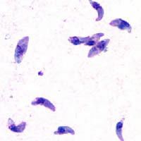
Tachyzoites
Motile, and quickly multiplying, tachyzoites are responsible for expanding the population of the parasite in the host. When a host consumes a tissue cyst (containing bradyzoites) or an oocyst (containing sporozoites), the bradyzoites or sporozoites stage-convert into tachyzoites upon infecting the intestinal epithelium of the host. During the initial, acute period of infection, tachyzoites spread throughout the body via the blood stream. During the later, latent (chronic) stages of infection, tachyzoites stage-convert to bradyzoites to form tissue cysts.
Merozoites
Like tachyzoites, merozoites divide quickly, and are responsible for expanding the population of the parasite inside the cat intestine prior to sexual reproduction. When a feline definitive host consumes a tissue cyst (containing bradyzoites), bradyzoites convert into merozoites inside intestinal epithelial cells. Following a brief period of rapid population growth in the intestinal epithelium, merozoites convert into the noninfectious sexual stages of the parasite to undergo sexual reproduction, eventually resulting in the formation of zygote-containing oocysts.
Bradyzoites
Bradyzoites are the slowly dividing stage of the parasite that make up tissue cysts. When an uninfected host consumes a tissue cyst, bradyzoites released from the cyst infect intestinal epithelial cells before converting to the proliferative tachyzoite stage. Following the initial period of proliferation throughout the host body, tachyzoites then convert back to bradyzoites, which reproduce inside host cells to form tissue cysts in the new host.
Sporozoites
Sporozoites are the stage of the parasite residing within oocysts. When a human or other warm-blooded host consumes an oocyst, sporozoites are released from it, infecting epithelial cells before converting to the proliferative tachyzoite stage.
Risk factors for human infection
The following have been identified as being risk factors for T. gondii infection:
Infection in humans and other warm-blooded animals can occur:
- by consuming raw or undercooked meat containing T. gondii tissue cysts. The most common threat to citizens in the United States is from eating raw or undercooked lamb or pork. It is possible, though unlikely, to ingest the parasite through other products:
- by ingesting water, soil, vegetables, or anything contaminated with oocysts shed in the feces of an infected animal. Cat fecal matter is particularly dangerous: Just one cyst consumed by a cat can result in thousands of oocysts. This is why physicians recommend pregnant or ill persons do not clean the cat's litter box at home. These oocysts are resilient to harsh environmental conditions and can survive over a year in contaminated soil.
- from a blood transfusion or organ transplant
- from transplacental transmission from mother to fetus, particularly when T. gondii is contracted during pregnancy
- from drinking unpasteurized goat milk
- by contact with soil
- from eating unwashed raw vegetables or fruits
Cleaning cat litter boxes is a potential route of infection; however, numerous studies have shown living in a household with a cat is not a significant risk factor for T. gondii infection, though living with several kittens has some significance.
In warm-blooded animals, such as brown rats, sheep, and dogs, T. gondii has also been shown to be sexually transmitted, and it is hypothesized that it may be sexually transmitted in humans, although not yet proven. Although T. gondii can infect, be transmitted by, and asexually reproduce within humans and virtually all other warm-blooded animals, the parasite can sexually reproduce only within the intestines of members of the cat family (felids). Felids are therefore defined as the definitive hosts of T. gondii, with all other hosts defined as intermediate hosts like human or other mammals.
Sewage has been identified as a carriage medium for the organism.
Preventing infection
The following precautions are recommended to prevent or greatly reduce the chances of becoming infected with T. gondii. This information has been adapted from the websites of United States Centers for Disease Control and Prevention and the Mayo Clinic.
From food
Basic food handling safety practices can prevent or reduce the chances of becoming infected with T. gondii, such as washing unwashed fruits and vegetable and avoiding raw or undercooked meat, poultry, and seafood. Other unsafe practices such as drinking unpasteurized milk or untreated water can increase odds of infection. Because T. gondii is typically transmitted through cysts that reside in the tissues of infected animals, meat that is not properly prepared can present an increased risk of infection. Freezing meat for several days at subzero temperatures (0 °F or −18 °C) before cooking eliminates tissue cysts, which can rarely survive these temperatures. During cooking, whole cuts of red meat should be cooked to an internal temperature of 145 °F (63 °C). Medium rare meat is generally cooked between 130 and 140 °F (55 and 60 °C), so cooking whole cuts of meat to medium is recommended. After cooking, a rest period of 3 min should be allowed before consumption. However, ground meat should be cooked to an internal temperature of at least 160 °F (71 °C) with no rest period. All poultry should be cooked to an internal temperature of at least 165 °F (74 °C). After cooking, a rest period of 3 min should be allowed before consumption.
From environment
Oocysts in cat feces take at least a day to sporulate and become infectious after they are shed, so disposing of cat litter daily greatly reduces the chances of infectious oocysts being present in litter. As infectious oocysts from cat feces can spread and survive in the environment for months, humans should wear gloves when gardening or working with soil, and should wash their hands promptly after disposing of cat litter. The same precautions apply to outdoor sandboxes, which should be covered when not in use.
Furthermore, pregnant or immunocompromised people are at higher risk of becoming infected or transmitting the parasite to their fetus. Because of this, they should not change or handle cat litter boxes. Ideally, cats should be kept indoors and only fed food that has low to no risk of carrying oocysts, such as commercial cat food or well-cooked table food.
Vaccination
As of 2016, no approved human vaccine exists against Toxoplasma gondii. Research on human vaccines is ongoing.
For sheep, an approved live vaccine sold as Toxovax (MSD Animal Health) provides lifetime protection.
History
In 1908, while working at the Pasteur Institute in Tunis, Charles Nicolle and Louis Manceaux discovered a protozoan organism in the tissues of a hamster-like rodent known as the gundi, Ctenodactylus gundi. Although Nicolle and Manceaux initially believed the organism to be a member of the genus Leishmania that they described as "Leishmania gondii", they soon realized they had discovered a new organism entirely. They named it Toxoplasma gondii, a reference to its morphology (Toxo, from Greek τόξον (toxon); arc, bow, and πλάσμα (plasma); i.e., anything shaped or molded) and the host in which it was discovered, the gundi (gondii). The same year Nicolle and Mancaeux discovered T. gondii, Alfonso Splendore identified the same organism in a rabbit in Brazil. However, he did not give it a name.
The first conclusive identification of T. gondii in humans was in an infant girl delivered full term by Caesarean section on May 23, 1938, at Babies' Hospital in New York City. The girl began having seizures at three days of age, and doctors identified lesions in the maculae of both of her eyes. When she died at one month of age, an autopsy was performed. Lesions discovered in her brain and eye tissue were found to have both free and intracellular T. gondii'. Infected tissue from the girl was homogenized and inoculated intracerebrally into rabbits and mice; the animals subsequently developed encephalitis. Later, congenital transmission was found to occur in numerous other species, particularly in sheep and rodents.
The possibility of T. gondii transmission via consumption of undercooked meat was first proposed by D. Weinman and A.H Chandler in 1954. In 1960, the cyst wall of tissue cysts was shown to dissolve in the proteolytic enzymes found in the stomach, releasing infectious bradyzoites into the stomach (and subsequently into the intestine). The hypothesis of transmission via consumption of undercooked meat was tested in an orphanage in Paris in 1965; yearly acquisition rates of T. gondii rose from 10% to 50% after adding two portions of barely cooked beef or horse meat to the orphans' daily diets, and to 100% after adding barely cooked lamb chops. Such an experiment would nowadays be considered unethical.
In 1959, a study in Bombay found the prevalence of T. gondii in strict vegetarians to be similar to that found in nonvegetarians. This raised the possibility of a third major route of infection, beyond congenital and carnivorous transmission. In 1970, the existence of oocysts was discovered in cat feces, and the fecal-oral route of infection via oocysts was demonstrated.
Throughout the 1970s and 1980s, a vast number of species were tested for the ability to shed oocysts upon infection. Whereas at least 17 different species of felids have been confirmed to shed oocysts, no nonfelid has been shown to be permissive for T. gondii sexual reproduction and subsequent oocyst shedding.
Behavioral differences of infected hosts
There are many instances where behavioural changes were reported in rodents with T. gondii. The changes seen were a reduction in their innate dislike of cats, which made it easier for cats to prey on the rodents. In an experiment conducted by Berdoy and colleagues the infected rats showed preference for the cat odour area versus the area with the rabbit scent, therefore making it easier for the parasite to take its final step in its definitive feline host. This is an example of the extended phenotype concept, that is, the idea that the behaviour of the infected animal changes in order to maximize survival of the genes that increase predation of the intermediate rodent host.
Differences in sex-dependent behavior observed in infected hosts compared to non-infected individuals can be attributed to differences in testosterone. Infected males had higher levels of testosterone while infected females had significantly lower levels, compared to their non-infected equivalents. Looking at humans, studies using the Cattell’s 16 Personality Factor questionnaire, found that infected men scored lower on Factor G (superego strength/rule consciousness) and higher on Factor L (vigilance) while the opposite pattern was observed for infected women. This means that men were more likely to disregard rule and were more expedient, suspicious and jealous. On the other hand, women were more warm hearted, outgoing, conscientious and moralistic. However, human studies have not been able to show causation as they have all been observational studies. Mice infected with T. gondii have a worse motor performance than non-infected mice. Thus, a computerized simple reaction test was given to both infected and non-infected adults. It was found that the infected adults performed much more poorly and lost their concentration more quickly than the control group. But, the effect of the infection only explains less than 10% of the variability in performance. (i.e., there could be other confounding factors) Correlation has also been observed between seroprevalence of T. gondii in humans and increased risk of traffic accidents. Infected subjects have a 2.65 times higher risk of getting into a traffic accident. A similar study done in Turkey showed that there is a higher incidence of Toxoplasma gondii antibodies among drivers who have been involved in traffic accidents. Furthermore, this parasite has been associated with many neurological disorders such as schizophrenia. In a meta-analysis, 23 studies met inclusion criteria. The results demonstrate that the seroprevalence of antibodies to T. gondii in people with schizophrenia is significantly higher than in control populations (OR=2.73, P<0.000001). More recent studies found that suicide attempters has significantly higher IgG antibody levels to T. gondii than patients without a suicide attempt. Infection was also shown to be associated with suicide in women over the age of 60. (P<0.005)
As mentioned before, these results of increased proportions of people seropositive for the parasite in cases of these neurological disorders do not necessarily indicate a causal relationship between the infection and disorder. It is also important to mention that in 2016 a population-representative birth cohort study which was done, to test a hypothesis that toxoplasmosis is related to impairment in brain and behaviour measured by a range of phenotypes including neuropsychiatric disorders, poor impulse control, personality and neurocognitive deficits. The results of this study did not support the results in the previously mentioned studies, more than marginally. None of the P-values showed significance for any outcome measure. Thus, according to this study, the presence of T. gondii antibodies is not correlated to increase susceptibility to any of the behaviour phenotypes (except possibly to a higher rate of unsuccessful attempted suicide). This team did not observe any significant association between T. gondii serpositivity and schizophrenia. The team notes that the null findings might be a false negative due to low statistical power because of small sample sizes, but against this weights that their set-up should avoid some possibilities for errors in the about 40 studies that did show a positive correlation. They conclude that further studies should be performed.