
Latin os sphenoidale TA A02.1.05.001 | MeSH A02.835.232.781.802 FMA 52736 | |
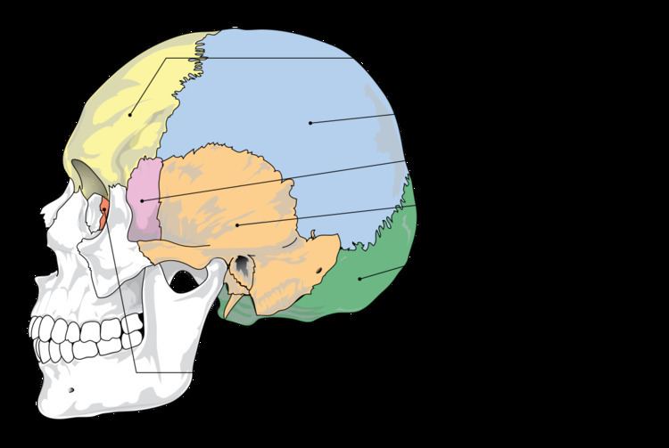 | ||
The sphenoid bone is an unpaired bone of the neurocranium. It is situated in the middle of the skull towards the front, in front of the temporal bone and the basilar part of the occipital bone. The sphenoid bone is one of the seven bones that articulate to form the orbit. Its shape somewhat resembles that of a butterfly or bat with its wings extended.
Contents
Structure
It is divided into the following parts:
Two sphenoidal conchae are situated at the anterior and inferior part of the body.
Intrinsic ligaments of the sphenoid
The more important of these are:
Note:These ligaments occasionally ossify.
Features
Articulations
The sphenoid articulates with the frontal, parietal, ethmoid, temporal, zygomatic, palatine, vomer, and occipital bones and helps to connect the neurocranium to the facial skeleton.
Superior or Cerebral Surface
Articulates with ethmoid bone anteriorly and basilar part of occipital bone posteriorly. It shows:
- Jugum sphenoidale
- Sulcus chiasmaticus
- Tuberculum sellae
- Sella turcica
- Dorsum sellae
- Clivus
Inferior surface
- Rostrum of sphenoid
- Sphenoidal conchae
- Vaginal processes of medial pterygoid plate
Anterior surface
Sphenoidal crest articulates with the perpendicular plate of ethmoid leading to formation of a part of the septum of nose.
Posterior surface
Basilar part of occipital bone
Lateral surface
Carotid sulcus lodging cavernous sinus and internal carotid artery
Sphenoidal sinuses
These are asymmetrical air sinuses in the body of the sphenoid, closed by Sphenoidal conchae
Superior or cerebral surface
This forms the floor of the middle cranial fossa. It presents (starting from the front):
Lateral surface
This is divided into (by infratemporal crest):
Foramen pierce it:
Orbital surface
This forms the posterior wall of the orbit
Lesser wings
These are two triangular wings projecting laterally from anterosuperior part of the body. Each consists of:
Development
Until the seventh or eighth month of fetal development, the body of the sphenoid consists of two parts: one in front of the tuberculum sellæ, the presphenoid, with which the small wings are continuous; the other, consisting of the sella turcica and dorsum sellae, the postsphenoid, with which are associated the great wings, and pterygoid processes.
The greater part of the bone is ossified in cartilage. There are fourteen centers in all, six for the presphenoid and eight for the postsphenoid.
Presphenoid
By about the ninth week of fetal development an ossific center appears for each of the small wings (orbito-sphenoids) just lateral to the optic foramen; this is followed by the appearance of two nuclei in the presphenoid part of the body.
The sphenoidal conchae are each developed from a center that makes its appearance about the fifth month; at birth they consist of small triangular laminae, and it is not until the third year that they become hollowed out and coneshaped; about the fourth year they fuse with the labyrinths of the ethmoid bone, and between the ninth and twelfth years they unite with the sphenoid bone.
Postsphenoid
The first ossific nuclei are those for the great wings (alisphenoids). One makes its appearance in each wing between the foramen rotundum and foramen ovale about the eighth week. The orbital plate and that part of the sphenoid, which is found in the temporal fossa, as well as the lateral pterygoid plate, are ossified in membrane (Fawcett).
Soon after, the centers for the postsphenoid part of the body appear, one on either side of the sella turcica, and become blended together about the middle of fetal life.
Each medial pterygoid plate (except its hamulus) is ossified in membrane, and its center probably appears about the ninth or tenth week; the hamulus becomes chondrified during the third month, and almost at once ossifies (Fawcett).
The medial joins the lateral pterygoid plate about the sixth month.
About the fourth month, a center appears for each lingula and speedily joins the rest of the bone.
The presphenoid is united to the postsphenoid about the eighth month, and at birth the sphenoid is in three pieces [Fig. 4]: a central, consisting of the body and small wings, and two lateral, each comprising a great wing and pterygoid process.
In the first year after birth the great wings and body unite, and the small wings extend inward above the anterior part of the body, and, meeting with each other in the middle line, form an elevated smooth surface, termed the jugum sphenoidale.
By the twenty-fifth year the sphenoid and occipital are completely fused.
Between the pre- and postsphenoid there are occasionally seen the remains of a canal, the canalis cranio-pharyngeus, through which, in early fetal life, the hypophyseal diverticulum of the buccal ectoderm is transmitted.
The sphenoidal sinuses are present as minute cavities at the time of birth (Onodi), but do not attain their full size until after puberty.
Function
This bone assists with the formation of the base and the sides of the skull, and the floors and walls of the orbits. It is the site of attachment for most of the muscles of mastication. Many foramina and fissures are located in the sphenoid that carry nerves and blood vessels of the head and neck, such as the superior orbital fissure (with ophthalmic nerve), foramen rotundum (with maxillary nerve) and foramen ovale (with mandibular nerve).
Other animals
The sphenoid bone of humans is homologous with a number of bones that are often separate in other animals, and have a somewhat complex arrangement.
In the early lobe-finned fishes and tetrapods, the pterygoid bones were flat, wing-like bones forming the major part of the roof of the mouth. Above the pterygoids were the epipterygoid bones, which formed part of a flexible joint between the braincase and the palatal region, as well as extending a vertical bar of bone towards the roof of the skull. Between the pterygoids lay an elongated, narrow parasphenoid bone, which also spread over some of the lower surface of the braincase, and connected, at its forward end, with a sphenethmoid bone helping to protect the olfactory nerves. Finally, the basisphenoid bone formed part of the floor of the braincase and lay immediately above the parasphenoid.
Aside from the loss of the flexible joint at the rear of the palate, this primitive pattern is broadly retained in reptiles, albeit with some individual modifications. In birds, the epipterygoids are absent and the pterygoids considerably reduced. Living amphibians have a relatively simplified skull in this region; a broad parasphenoid forms the floor of the braincase, the pterygoids are relatively small, and all other related bones except the sphenethmoid are absent.
In mammals, these various bones are often (though not always) fused into a single structure; the sphenoid. The basisphenoid forms the posterior part of the base, while the pterygoid processes represent the pterygoid bones. The epipterygoids have extended into the wall of the cranium; they are referred to as alisphenoids when separate in mammals, and form the greater wings of the sphenoid when fused into a larger structure. The sphenethmoid bone forms as three bones: the lesser wings and the anterior part of the base. These two parts of the sphenethmoid may be distinguished as orbitosphenoids and presphenoid, respectively, although there is often some degree of fusion. Only the parasphenoid appears to be entirely absent in mammals.
In the dog the sphenoid is represented by 8 bones: basisphenoid, alisphenoids, presphenoid, orbitosphenoids, pterygoids. These bones remain separate and are the: