
From trigeminal nerve TA A14.2.01.016 | Latin n. ophthalmicus FMA 52621 | |
 | ||
MeSH A08.800.800.120.760.650 | ||
The ophthalmic nerve (CN V: V1) is one of the three branches of the trigeminal nerve, the fifth cranial nerve. It carries sensory information from the face/scalp and sympathetic fibers for pupil dilation in the long ciliary branch of the nasociliary nerve.
Contents
Structure
The frontal branch passes through the orbit superiorly, then reenters the frontal bone briefly before exiting above the orbit through the superior orbital fissure and the supraorbital notch to provide sensory innervation for the skin of the forehead and scalp. The lacrimal nerve passes through the orbit superiorly to innervate the lacrimal gland. The nasociliary branch gives off several sensory branches to the orbit and then continues out through the anterior ethmoidal foramen, where it enters the nasal cavity and provides innervation for much of the anterior nasal mucosa. It also gives off a branch which exits through the nasal bones to form the external nasal branch.
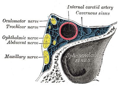
The ophthalmic nerve is joined by filaments from the cavernous plexus of the sympathetic, and communicates with the oculomotor, trochlear, and abducent nerves; it gives off a recurrent (meningeal) filament which passes between the layers of the tentorium.
Branches
Function
The ophthalmic nerve supplies branches to the cornea, ciliary body, and iris; to the lacrimal gland and conjunctiva; to the part of the mucous membrane of the nasal cavity; and to the skin of the eyelids, eyebrow, forehead and nose.
It is the smallest of the three divisions of the trigeminal, and arises from the upper part of the trigeminal ganglion as a short, flattened band, about 2.5 cm. long, which passes forward along the lateral wall of the cavernous sinus, below the oculomotor and trochlear nerves; just before entering the orbit, through the superior orbital fissure, it divides into three branches, lacrimal, frontal, and nasociliary.
It carries sensory branches from the eyes, conjunctiva, lacrimal gland, nasal cavity, frontal sinus, ethmoidal cells, falx cerebri, dura mater in the anterior cranial fossa,superior parts of the tentorium cerebelli, upper eyelid, dorsum of the nose, and anterior part of the scalp.
Roughly speaking, the ophthalmic nerve supplies general somatic afferents to the upper face, skull, and eye:
Compare this to the maxillary nerve, which supplies general somatic afferents to the mid-face and skull: