
Dorlands/Elsevier f_08/12365608 | FMA 54799 | |
 | ||
Latin fissura orbitalis superior TA A02.1.05.023A02.1.00.083 | ||
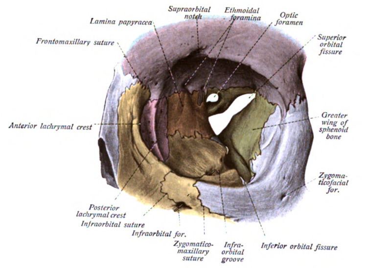
The superior orbital fissure is a foramen in the skull, although strictly it is more of a cleft, lying between the lesser and greater wings of the sphenoid bone.
Contents
Structures passing through
A number of important anatomical structures pass through the fissure, and these can be damaged in orbital trauma, particularly blowout fractures through the floor of the orbit into the maxillary sinus. These structures are:
These include nonvisual sensory messages, such as pain, or motor nerves. They also serve as vascular connections.
The nerves passing through the fissure can be remembered with the mnemonic, "Live Frankly To See Absolutely No Insult" - for Lacrimal and Frontal divisions of the ophthalmic nerve (V1), Trochlear nerve (IV), Superior division of the oculomotor nerve (III), Abducens nerve (VI), Nasociliary branch of the ophthalmic nerve (V1) and Inferior Division of the oculomotor nerve (III).
It is divided into 3 parts from lateral to medial:
Pathology
The abducens nerve is most likely to show signs of damage first, with the most common complaints retro-orbital pain and the involvement of cranial nerves III, IV, V1, and VI without other neurological signs or symptoms. This presentation indicates either compression of structures in the superior orbital fissure or the cavernous sinus.
Superior orbital fissure syndrome
Superior orbital fissure syndrome, also known as Rochon-Duvigneaud's syndrome, is a neurological disorder that results if the superior orbital fissure is fractured. Involvement of the cranial nerves that pass through the superior orbital fissure may lead to diplopia, paralysis of extraocular muscles, exophthalmos, and ptosis. Blindness or loss of vision indicates involvement of the orbital apex, which is more serious, requiring urgent surgical intervention. Typically, if blindness is present with superior orbital syndrome, it is called orbital apex syndrome.