
Latin Foramen spinosum TA A02.1.05.038 | Dorlands/Elsevier f_12/12373713 FMA 53156 | |
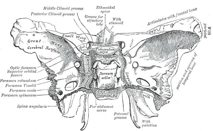 | ||
The foramen spinosum is one of two foramina located in the base of the human skull, on the sphenoid bone. It is situated just anterior to the spine of the sphenoid bone, and just lateral to the foramen ovale. The middle meningeal artery, middle meningeal vein, and the meningeal branch of the mandibular nerve pass through the foramen.
Contents
The foramen spinosum is often used as a landmark in neurosurgery, due to its close relations with other cranial foramina. It was first described by Jakob Benignus Winslow in the 18th century.
Structure
The foramen spinosum is a foramen through the sphenoid bone situated in the middle cranial fossa. It is one of two foramina in the greater wing of the sphenoid bone. The foramen ovale is one of these two cranial foramina, situated directly anterior and medial to the foramen spinosum. The spine of sphenoid falls medial and posterior to the foramen. Lateral to the foramen is the mandibular fossa, and posterior is the Eustachian tube.
Variation
The foramen spinosum varies in size and location. The foramen is rarely absent, usually unilaterally, in which case the middle meningeal artery enters the cranial cavity through the foramen ovale. It may be incomplete, which may occur in almost half of the population. Conversely, in a minority of cases (less than 1%), it may also be duplicated, particularly when the middle meningeal artery is also duplicated.
The foramen may pass through the sphenoid bone at the apex of the spinous process, or along its medial surface.
Development
In the newborn, the foramen spinosum is about 2.25 mm long and in adults about 2.56 mm. The width of the foramen extends from 1.05 mm to about 2.1 mm in adults. The average diameter of the foramen spinosum is 2.63 mm in adults.
The earliest perfect ring-shaped formation of the foramen spinosum was observed in the eighth month after birth and the latest seven years after birth in a developmental study of the foramen rotundum, foramen ovale and foramen spinosum. The majority of the foramina in the skull studies were round in shape. The sphenomandibular ligament, derived from the first pharyngeal arch and usually attached to the spine of the sphenoid bone, may be found attached to the rim of the foramen.
Animals
In other great apes, the foramen spinosum is found not in the sphenoid bone but in parts of the temporal bone such as the squamous part, found at the sphenosquamosal suture, or absent.
Function
The foramen spinosum permits the passage of the middle meningeal artery, middle meningeal vein, and the meningeal branch of the mandibular nerve.
Clinical significance
Due to its distinctive position, the foramen is used as an anatomical landmark during neurosurgery. As a landmark, the foramen spinosum reveals the positions of other cranial foramina, the mandibular nerve and trigeminal ganglion, foramen ovale, and foramen rotundum. It may also be relevant in achieving haemostasis during trauma surgery.
History
The foramen spinosum was first described by the Danish anatomist Jakob Benignus Winslow in the 18th century. It is so-named because of its relationship to the spinous process of the greater wing of the sphenoid bone. However, due to incorrectly declining the noun, the literal meaning is "hole full of thorns" (Latin: foramen spinosum). The correct, but unused name would, in fact, be foramen spinae.