
Group Group V ((−)ssRNA) Scientific name Rhabdoviridae | Order Mononegavirales Rank Family | |
 | ||
Lower classifications Lyssavirus, Infectious hematopoietic necrosis, Chandipura virus, Rabies virus, Vesicular stomatitis virus | ||
Rhabdoviridae
Rhabdoviridae is a family of virus in the order Mononegavirales. Vertebrates (including mammals and humans), invertebrates, and plants serve as natural hosts. There are currently 74 species in this family, divided among 11 genera. Diseases associated with viruses of this family include rabies fatal encephalitis caused by rabies virus, and vesicular diseases and encephalitis flu-like symptoms in humans caused by vesiculoviruses. The name is derived from the Greek rhabdos meaning rod referring to the shape of the viral particles.
Contents
- Rhabdoviridae
- Micro vet 2015 2 aula 7 fam lia rhabdoviridae
- Structure
- Taxonomy
- Transcription
- Translation
- Replication
- Clades
- Proposed Classifications
- Prototypical Rhabdoviruses
- References
Micro vet 2015 2 aula 7 fam lia rhabdoviridae
Structure
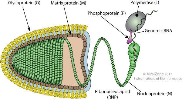
Rhabdovirions are enveloped, with bullet shaped and bacilliform geometries. These virions are about 75 nm wide and 180 nm long. Rhabdoviruses have helical nucleocapsids and their genomes are linear, around 11–15 kb in length. Rhabdoviruses carry their genetic material in the form of negative-sense single-stranded RNA. They typically carry genes for five proteins: large protein (L), glycoprotein (G), nucleoprotein (N), phosphoprotein (P), and matrix protein (M). Rhabdoviruses that infect vertebrates( especially mammals and fishes), plants and insects are usually bullet-shaped., However, in contrast to paramyxoviruses, rhabdoviruses do not have hemagglutinating and neuraminidase activities.
Taxonomy
Table legend: "*" denotes type species.
In addition to the above, there are a large number of rhabdo-like viruses (~130) that have not yet been officially classified by the ICTV.
Transcription
Transcriptase of rhabdovirus is composed of 1 L and 3 P proteins. Transcriptase components always presents in the virion; so rhabdoviruses can start transcription right after the entry with no need to produce anything.
Transcriptase starts to move from 3' end to 5' end on the genome, and the movement terminates randomly at the end of protein sequences. For example, if a transcription finishes at the end of M sequence; leader RNA and N, P and M mRNAs are formed separately from each other.
Also mRNAs accumulate according to the order of protein sequences on the genome, and this solves the logistics problem in the cell. For example, N protein is needed too much for the virus, because it coats outside of the replicated genomes completely. Because of having the N protein sequence at the beginning of the genome (3' end) after the leader RNA sequence, mRNAs for N protein can always be produced and accumulate in high amounts with every termination of transcription. After the transcription processes, all of the mRNAs are capped at the 5' end and polyadenylated at the 3' end by L protein.
This transcription mechanism provides to produce mRNAs according to the need of the viruses.
Translation
The virus proteins translated on free ribosomes but G protein is translated by the rough endoplasmic reticulum. This means G protein has a signal peptide on its mRNA's starting codes. Phosphoproteins(P) and glycoprotein(G) undergo post-translational modification. Trimers of P protein are formed after phosphorylation by kinase activity of L protein. The G protein is glycosylated in the rough endoplasmic reticulum and the Golgi complex.
Replication
Viral replication is cytoplasmic. Entry into the host cell is achieved by attachment of the viral G glycoproteins to host receptors, which mediates clathrin-mediated endocytosis. Replication follows the negative stranded RNA virus replication model. Negative stranded RNA virus transcription, using polymerase stuttering is the method of transcription. The virus exits the host cell by budding, and tubule-guided viral movement. Transmission routes are zoonosis and bite.
Replication of many rhabdoviruses occurs in the cytoplasm, although several of the plant infecting viruses replicate in the nucleus.. In order for replication, both the L and P protein must be expressed to regulate transcription. The L protein have a lot of enzymatic actiivites such as RNA replication, capping mRNAs phospholorylation of P. L protein gives feature in about replication in cytopolasm. Transcription results in five monocistronic mRNAs being produced because the intergenic sequences act as both termination and promoter sequences for adjacent genes. This type of transcription mechanism is explained by stop-start model( stuttering transcription). Owing to stop-start model, the large amounts of the structural proteins are produced. According to this model, the virus-associated RNA polymerase starts firstly the synthesis of leader RNA and then the five mRNA which will produce N,P,M,G,L proteins, respectively. After the leader RNA was produced, the polymerase enzyme reinitiates virion transcription on N gene and proceeds its synthesis until it ends 3′ end of the chain.Then, the synthesis of P mRNAs are made by same enzyme with new starter sinyal. These steps continue until the enzyme arrives the end of the L gene. During transcription process, the polymerase enzyme may leave the template at any point and then bound just at the 3′ end of the genome RNA to start mRNA synthesis again. This process will results concentration gradient of the amount of mRNA based on its place and its range from the 3′ end. In the circumstances, the amounts of mRNA species change and will be produced N>P>M>G>L proteins. During their synthesis the mRNAs are processed to introduce a 5' cap and a 3’ polyadenylated tail to each of the molecules .This structure is homologous to cellular mRNAs and can thus be translated by cellular ribosomes to produce both structural and non-structural proteins.
Genomic replication requires a source of newly synthesized N protein to encapsidate the RNA. This occurs during its synthesis and results in the production of a full length anti-genomic copy. This in turn is used to produce more negative-sense genomic RNA. The viral polymerase is required for this process, but how the polymerase engages in both mRNA synthesis and genomic replication is not well understood.
Replication characteristically occurs in an inclusion body within the cytoplasm, from where they bud through various cytoplasmic membranes and the outer membrane of the cell. This process results in the acquisition of the M + G proteins, responsible for the characteristic bullet- shaped morphology of the virus.
Clades
These viruses fall into four groups based on the RNA polymerase gene. The basal clade appears to be novirhabdoviruses, which infect fish. Cytorhabdoviruses and the nucleorhabdoviruses, which infect plants, are sister clades. Lyssaviruses form a clade of their own which is more closely related to the land vertebrate and insect clades than to the plant viruses. The remaining viruses form a number of highly branched clades and infect arthropods and land vertebrates.
A 2015 analysis of 99 species of animal rhabdoviruses found that they fell into 17 taxonomic groupings, eight – Lyssavirus, Vesiculovirus, Perhabdovirus, Sigmavirus, Ephemerovirus, Tibrovirus, Tupavirus and Sprivivirus' - which were previously recognized. The authors proposed seven new taxa on the basis of their findings: "Almendravirus", "Bahiavirus", "Curiovirus", "Hapavirus", "Ledantevirus", "Sawgravirus" and "Sripuvirus". Seven species did not group with the others suggesting the need for additional taxa.
Proposed Classifications
"Curioviruses" a group of four viruses that were isolated from biting midges (Culicoides), sandflies (Lutzomyia ) and mosquitoes (Coqillettidia and Trichoprosopon) which were captured in the forests of South America and the Caribbean.
"Bracorhabdoviruses" are derived from the acronym Brazilian Amazonian Culicoides rhabdoviruses.
An inofficial supergroup – "Dimarhabdovirus" – refers to the genera Ephemerovirus and Vesiculovirus. A number of other viruses that have not been classified into genera also belong to this taxon. This supergroup contains the genera with species that replicate in both vertebrate and invertebrate hosts and have biological cycles that involve transmission by haematophagous dipterans.
Prototypical Rhabdoviruses
The prototypical and best studied rhabdovirus is vesicular stomatitis Indiana virus. It is a preferred model system to study the biology of rhabdoviruses, and mononegaviruses in general.
The mammalian disease rabies is caused by lyssaviruses, of which several have been identified.
Rhabdoviruses are important pathogens of animals and plants. Rhabdoviruses are transmitted to hosts by arthropods, such as aphids, planthoppers, leafhoppers, black flies, sandflies, and mosquitoes.
In September 2012, researchers writing in the journal PLOS Pathogens described a species of rhabdovirus, called Bas-Congo Virus or BASV, responsible for 4 cases of viral hemorrhagic fever in the Bas-Congo district in 2009. The 2 non-fatal cases occurred in healthcare workers treating the other 2, suggesting the possibility of person-to-person transmission.