
Specialty infectious disease DiseasesDB 33989 | ICD-10 B88.8 (ILDS B88.83) eMedicine derm/348 | |
 | ||
Protothecosis is a disease found in dogs, cats, cattle, and humans caused by a type of green alga known as Prototheca that lacks chlorophyll. It and its close relative Helicosporidium are unusual in that they are actually green algae that have become parasites. The two most common species are Prototheca wickerhamii and Prototheca zopfii. Both are known to cause disease in dogs, while most human cases are caused by P. wickerhami. Prototheca is found worldwide in sewage and soil. Infection is rare despite high exposure, and can be related to a defective immune system. In dogs, females and Collies are most commonly affected.
Contents
The first human case was identified in 1964 in Sierra Leone.
Treatment
Treatment with amphotericin B has been reported.
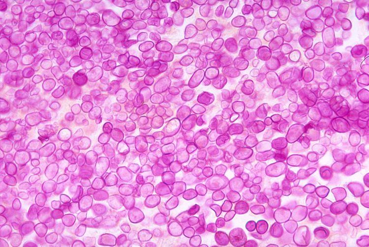
The organism
Prototheca has been thought to be a mutant of Chlorella, a type of single-celled green alga. However, while Chlorella contains galactose and galactosamine in the cell wall, Prototheca lacks these. Also, Chlorella obtains its energy through photosynthesis, while Prototheca is saprotrophic, feeding on dead and decaying organic matter. When Prototheca was first isolated from slime flux of trees in 1894, it was thought to be a type of fungus. Its size varies from 2 to 15 micrometres.
Protothecosis in cattle
Cattle can be affected by protothecal enteritis and mastitis. Protothecal mastitis is endemic worldwide, although most cases of infected herds have been reported in Germany, the United States, and Brazil.
Protothecosis in dogs
Disseminated protothecosis is most commonly seen in dogs. The algae enters the body through the mouth or nose and causes infection in the intestines. From there it can spread to the eye, brain, and kidneys. Symptoms can include diarrhea, weight loss, weakness, inflammation of the eye (uveitis), retinal detachment, ataxia, and seizures.
Dogs with acute blindness and diarrhea that develop exudative retinal detachment should be assessed for protothecosis. Diagnosis is through culture or finding the organism in a biopsy, cerebrospinal fluid, vitreous humour, or urine. Treatment of the disseminated form in dogs is very difficult, although use of antifungal medication has been successful in a few cases. Prognosis for cutaneous protothecosis is guarded and depends on the surgical options. Prognosis for the disseminated form is grave. This may be due to delayed recognition and treatment.