
Specialty dermatology ICD-9-CM 694.4 MedlinePlus 000882 | ICD-10 L10 DiseasesDB 9764 3117933489 | |
 | ||
OMIM 169600 Benign Chronic ~169610 ~ Vulgaris, Familial | ||
Pemphigus (/ˈpɛmfɪɡəs/ or /pɛmˈfaɪɡəs/) is a rare group of blistering autoimmune diseases that affect the skin and mucous membranes. The name is derived from the Greek root "pemphix" meaning "pustule."
Contents
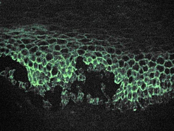
In pemphigus, autoantibodies form against desmoglein. Desmoglein forms the "glue" that attaches adjacent epidermal cells via attachment points called desmosomes. When autoantibodies attack desmogleins, the cells become separated from each other and the epidermis becomes "unglued", a phenomenon called acantholysis. This causes blisters that slough off and turn into sores. In some cases, these blisters can cover a significant area of the skin.
Originally, the cause of this disease was unknown, and "pemphigus" was used to refer to any blistering disease of the skin and mucosa. In 1964, researchers found that the blood of patients with pemphigus contained antibodies to the layers of skin that separate to form the blisters. In 1971, an article investigating the autoimmune nature of this disease was published.
Types
There are several types of pemphigus which vary in severity: pemphigus vulgaris, pemphigus foliaceus, Intraepidermal neutrophilic IgA dermatosis, and paraneoplastic pemphigus.
Note that Hailey-Hailey disease, also called familial benign pemphigus, is an inherited (genetic) skin disease, not an autoimmune disease. It is therefore not considered part of the Pemphigus group of diseases.
Diagnosis
Pemphigus defines a group of autoimmune interepithelial blistering diseases that are characterized by loss of normal cell-cell adhesion (acantholysis), and by the presence of pathogenic (predominantly IgG) autoantibodies reacting against epithelial adhesion molecules. Pemphigus is further divided in two major subtypes: pemphigus vulgaris (PV) and pemphigus foliaceus (PF). However, several other disorders such as IgA pemphigus, IgE pemphigus, pemphigus herpetiformis, drug induced pemphigus, Senear Usher syndrome and endemic pemphigus foliaceus exist;recognized by a dermatologist from the appearance and distribution of the skin lesions. It is also commonly diagnosed by specialists practicing otolaryngology- head and neck surgery, periodontists, oral and maxillofacial surgeons and eye doctors, as lesions can affect the eyes and mucous membrane of the oral cavity. Intraorally it resembles the more common diseases lichen planus and mucous membrane pemphigoid. Definitive diagnosis requires examination of a skin or mucous membrane biopsy by a dermatopathologist or oral pathologist. The skin biopsy is taken from the edge of a blister, prepared for histopathology and examined with a microscope. The pathologist looks for an intraepidermal vesicle caused by the breaking apart of epidermal cells (acantholysis). Thus, the superficial (upper) portion of the epidermis sloughs off, leaving the bottom layer of cells on the "floor" of the blister. This bottom layer of cells is said to have a "tombstone appearance".
Definitive diagnosis also requires the demonstration of anti-desmoglein autoantibodies by direct immunofluorescence on the skin biopsy. These antibodies appear as IgG deposits along the desmosomes between epidermal cells, a pattern reminiscent of chicken wire. Anti-desmoglein antibodies can also be detected in a blood sample using the ELISA technique.
Classification
Pemphigus is a group of autoimmune blistering diseases that may be classified into the following types:
Treatment
If not treated, pemphigus can be fatal, usually from overwhelming opportunistic infection of lesions. The most common treatment is the administration of oral steroids, especially prednisone, often in high doses. The side effects of corticosteroids may require the use of so-called steroid-sparing or adjuvant drugs. One of the most dangerous side effects of high dosage steroid treatments is intestinal perforations, which may lead to sepsis. Steroids and other medications being taken to treat Pemphigus may also mask the effects of the perforations. Patients on high dosages of oral steroids should closely monitor their GI health. As lesions are usually terribly painful, it is likely that pain medication can complicate and exacerbate the GI issues caused by steroids.
Treatment Options
All of these drugs may cause severe side effects, so the patient should be closely monitored by doctors. Once the outbreaks are under control, dosage is often reduced, to lessen side effects.
If skin lesions do become infected, antibiotics may be prescribed. Tetracycline antibiotics have a mildly beneficial effect on the disease and are sometimes enough for Pemphigus Foliaceus. In addition, talcum powder is helpful to prevent oozing sores from adhering to bedsheets and clothes. Wound care and treatment is often akin to that used in burn units, including careful use of dressings that don't stick to the wounds, etc.
If paraneoplastic pemphigus is diagnosed with pulmonary disease, a powerful cocktail of immune suppressant drugs is sometimes used in an attempt to halt the rapid progression of bronchiolitis obliterans, including methylprednisolone, ciclosporin, azathioprine, and thalidomide. Plasmapheresis may also be useful.
Animals Affected
Pemphigus foliaceus has been recognized in pet dogs, cats, and horses and is the most common autoimmune skin disease diagnosed in veterinary medicine. Pemphigus foliaceus in animals produces clusters of small vesicles that quickly evolve into pustules. Pustules may rupture, forming erosions or become crusted. Left untreated, pemphigus foliaceus in animals is life-threatening, leading to not only loss of condition but also secondary infection.
Pemphigus vulgaris is a very rare disorder described in pet dogs and cats. Paraneoplastic pemphigus has been identified in pet dogs.