
Specialty oncology ICD-9-CM 212.7 OMIM 255960 | ICD-10 D21.9 ICD-O M8840/0 DiseasesDB 30736 | |
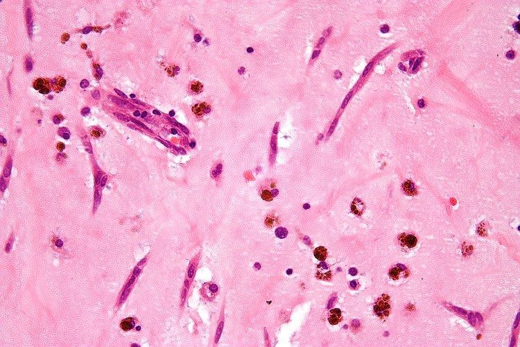 | ||
A myxoma (New Latin from Greek 'muxa' for mucus) is a myxoid tumor of primitive connective tissue. It is the most common primary tumor of the heart in adults, but can also occur in other locations.
Contents
Location
Atrial myxoma
Myxomas are usually located in either the left or right atrium of the heart; about 86 percent occur in the left atrium.
Myxomas are typically pedunculated, with a stalk that is attached to the interatrial septum. The most common location for attachment of the stalk is the fossa ovalis region of the interatrial septum.
An atrial myxoma may create an extra heart sound, audible to auscultation just after S2 It is most seen on echocardiography, as a pedunculated mass that is heterogeneous in appearance. A left atrial myxoma will cause an increase in pulmonary capillary wedge pressure.
The differential diagnosis include other cardiac tumors such as lipomas and rhabdomyomas (and rarely teratomas). These other tumors of the heart are typically not pedunculated, however, and are more likely to infiltrate the muscle of the heart. Cardiac magnetic resonance imaging (MRI) can help non-invasively diagnose cardiac tumors. However, diagnosis usually requires examination of a tissue sample by a pathologist.
Types
1.^ SMA, smooth muscle actin. 2.^ MSA, muscle-specific actin. 3.^ EMA, epithelial membrane antigen.
Symptoms
Symptoms associated with cardiac myxomas are typically due to the effect of the mass of the tumor obstructing the normal flow of blood within the chambers of the heart. Because pedunculated myxomas are somewhat mobile, symptoms may only occur when the patient is in a particular position.
Some symptoms of myxoma may be associated with the release of interleukin 6 (IL-6) by the myxoma. High levels of IL-6 may be associated with a higher risk of embolism of the myxoma.
Symptoms of a cardiac myxoma include:
Treatment
Myxomas are usually removed surgically. The surgeon removes the myxoma, along with at least 5 surrounding millimeters of atrial septum. The septum is then repaired, using material from the pericardium.