
Nerve Phrenic nerve Greek περίκάρδιον TA A12.1.08.001 | Latin Pericardium MeSH A07.541.795 | |
 | ||
Artery Pericardiacophrenic artery | ||
The pericardium (from the Greek περί, "around" and κάρδιον, "heart") is a double-walled sac containing the heart and the roots of the great vessels. The pericardial sac has two layers, a serous layer and a fibrous layer. It encloses the pericardial cavity which contains pericardial fluid.
Contents
- Structure
- Fibrous pericardium
- Serous pericardium
- Anatomical relationships
- Functions
- Clinical significance
- References
The pericardium fixes the heart to the mediastinum, gives protection against infection, and provides the lubrication for the heart.
Structure
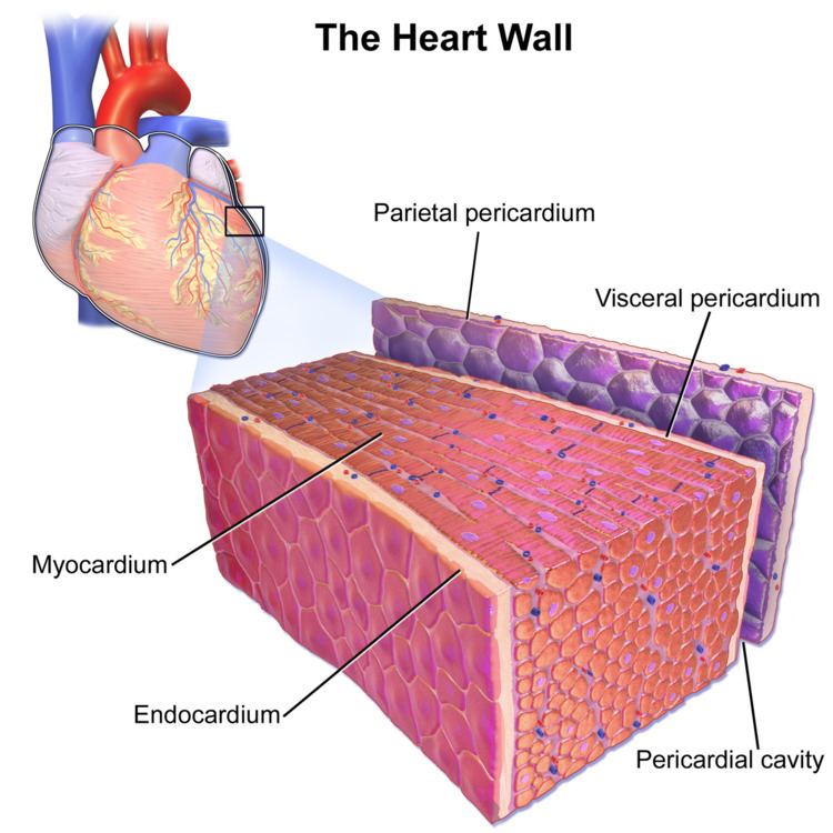
The pericardium is a tough double layered fibroserous sac which covers the heart. The space between the two layers of serous pericardium (see below), the pericardial cavity, is filled with serous fluid which protects the heart from any kind of external jerk or shock. There are two layers to the pericardial sac: the outermost fibrous pericardium and the inner serous pericardium.
Fibrous pericardium
The fibrous pericardium is the most superficial layer of the pericardium. It is made up of dense and loose connective tissue, which acts to protect the heart, anchoring it to the surrounding walls, and preventing it from overfilling with blood. It is continuous with the outer adventitial layer of the neighboring great blood vessels. It is inside the great blood vessels.
Serous pericardium
The serous pericardium, in turn, is divided into two layers, the parietal pericardium, which is fused to and inseparable from the fibrous pericardium, and the visceral pericardium, which is part of the epicardium. Both of these layers function in lubricating the heart to prevent friction during heart activity.
The visceral layer extends to the beginning of the great vessels (the large blood vessels serving the heart) becoming one with the parietal layer of the serous pericardium. This happens at two areas: where the aorta and pulmonary trunk leave the heart and where the superior vena cava, inferior vena cava and pulmonary veins enter the heart.
In between the parietal and visceral pericardial layers there is a potential space called the pericardial cavity, which contains a supply of lubricating serous fluid known as the pericardial fluid.
When the visceral layer of serous pericardium comes into contact with heart (not the great vessels) it is known as the epicardium. The epicardium is the layer immediately outside of the heart muscle proper (the myocardium). The epicardium is largely made of connective tissue and functions as a protective layer. During ventricular contraction, the wave of depolarization moves from the endocardial to the epicardial surface.
Anatomical relationships
Functions
Clinical significance
Inflammation of the pericardium is called pericarditis, and may be detected by the medical sign pericardial friction rub.
An excess of fluid in the cavity (such as in a pericardial effusion) can result in cardiac tamponade (compression of the heart within the pericardial sac). A pericardiectomy is sometimes needed to remedy this. The surgical removing of the epicardium is referred to as epicardiectomy.