
EC number 2.1.1.13 | Entrez 4548 | |
 | ||
External IDs OMIM: 156570 MGI: 894292 HomoloGene: 37280 GeneCards: MTR | ||
Methionine synthase also known as MS, MeSe, MetH is responsible for the regeneration of methionine from homocysteine. In humans it is encoded by the MTR gene (5-methyltetrahydrofolate-homocysteine methyltransferase). Methionine synthase forms part of the S-adenosylmethionine (SAMe) biosynthesis and regeneration cycle. In animals this enzyme requires Vitamin B12 (cobalamin) as a cofactor, whereas the form found in plants is cobalamin-independent. Microorganisms express both cobalamin-dependent and cobalamin-independent forms.
Contents
Mechanism
Methionine synthase catalyzes the final step in the regeneration of methionine(Met) from homocysteine(Hcy). The overall reaction transforms 5-methyltetrahydrofolate(N5-MeTHF) into tetrahydrofolate (THF) while transferring a methyl group to Hcy to form Met. Methionine synthase is the only mammalian enzyme that metabolizes N5-MeTHF to regenerate the active cofactor THF. In cobalamin-dependent forms of the enzyme, the reaction proceeds by two steps in a ping-pong reaction. The enzyme is initially primed into a reactive state by the transfer of a methyl group from N5-MeTHF to Co(I) in enzyme-bound cobalamin(Cob), forming methyl-cobalamin(Me-Cob) that now contains Me-Co(III) and activating the enzyme. Then, a Hcy that has coordinated to an enzyme-bound zinc to form a reactive thiolate reacts with the Me-Cob. The activated methyl group is transferred from Me-Cob to the Hcy thiolate, which regenerates Co(I) in cob, and Met is released from the enzyme. The cob-independent mechanism follows the same general pathway but with a direct reaction between the zinc thiolate and N5-MeTHF.
The mechanism of the enzyme depends on the constant regeneration of Co(I) in cob, but this is not always guaranteed. Instead, every 1-2000 catalytic turnovers, the Co(I) may be oxidized into Co(II), which would permanently shut down catalytic activity. A separate protein, Methionine Synthase Reductase, catalyzes the regeneration of Co(I) and the restoration of enzymatic activity. Because the oxidation of cob-Co(I) inevitably shuts down cob-dependent methionine synthase activity, defects or deficiencies in methionine synthase reductase have been implicated in some of the disease associations for methionine synthase deficiency discussed below. The two enzymes form a scavenger network seen on the lower left.
Structure
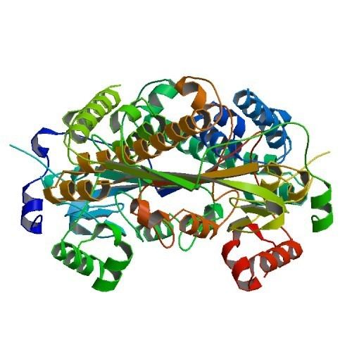
Crystal structures for both cob-independent and cob-dependent MetH have been solved, with little similarity in the overall structure despite the identical net reaction being performed by each and similarities within binding sites such as Hcy binding site. Cob-dependent MetH is divided into 4 separate domains: Activation, Cobalamin-binding(Cob domain), Homocysteine binding(Hcy domain), and N 5-methylTHF binding(MeTHF domain). The activation domain is the site of interaction with Methionine Synthase Reductase and binds SAM that is used as part of the re-activation cycle of the enzyme. The Cob domain contains Cob sandwiched between several large alpha helices and bound to the enzyme so that the cobalt atom of the group is exposed for contact with other domains. The Hcy domain contains the critical zinc-binding site, made up of cysteine or histidine residues coordinated to a zinc ion that can bind Hcy, with an example from a non-Cob dependent MetH shown on the right. The N5-MeTHF binding domain contains a conserved barrel in which N5-MeTHF can hydrogen bond with asparagine, arginine, and aspartic acid residues. The entire structure undergoes a dramatic swinging motion during catalysis as the Cob domain moves back and forth from the Hcy domain to the Fol domain, transferring the active methyl group from the Fol to Hcy domain.
Function
Methionine synthase's main purpose is to regenerate Met in the S-Adenosyl Methionine cycle, which in a single turnover consumes Met and ATP and generates Hcy. This cycle is critical because S-adenosyl methionine is used extensively in biology as a source of an active methyl group, and so methionine synthase serves an essential function by allowing the SAM cycle to perpetuate without a constant influx of Met. In this way, methionine synthase also serves to maintain low levels of Hcy and, because methionine synthase is one of the few enzymes that used N5-MeTHF as a substrate, to indirectly maintain THF levels.
In plants and microorganisms, methionine synthase serves a dual purpose of both perpetuating the SAM cycle and catalyzing the final synthetic step in the de novo synthesis of Met. While the reaction is exactly the same for both processes, the overall function is distinct from methionine synthase in humans because Met is an essential amino acid that is not synthesized de novo in the body.
Clinical significance
Mutations in the MTR gene have been identified as the underlying cause of methylcobalamin deficiency complementation group G, or methylcobalamin deficiency cblG-type. Deficiency or deregulation of the enzyme due to deficient methionine synthase reductase can directly result in elevated levels of homocysteine, which is associated with blindness, neurological symptoms, and birth defects. Most cases of methionine synthase deficiency are symptomatic within 2 years of birth with many patients rapidly developing severe encephalopathy. One consequence of reduced methionine synthase activity that is measurable by routine clinical blood tests is megaloblastic anemia.
Genetics
Several polymorphisms in the MTR gene have been identified.